

The IntI1 integron integrase preferentially binds single-stranded DNA of the attC site
- PMID: 10542191
- PMCID: PMC94154
- DOI: 10.1128/JB.181.21.6844-6849.1999
The IntI1 integron integrase preferentially binds single-stranded DNA of the attC site
Abstract
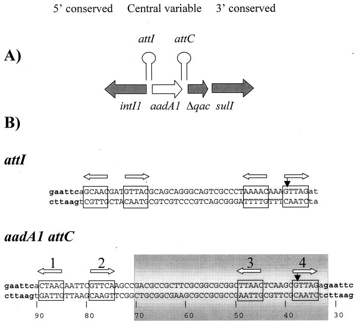
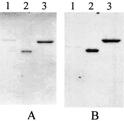
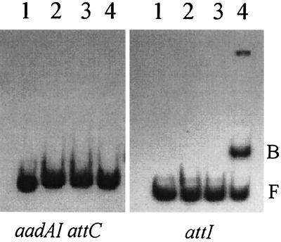
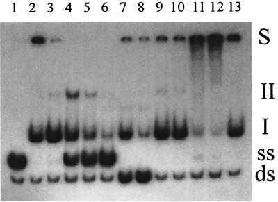
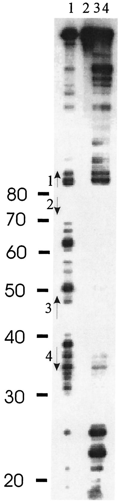
IntI1 integrase is a member of the prokaryotic DNA integrase superfamily. It is responsible for mobility of antibiotic resistance cassettes found in integrons. IntI1 protein, as well as IntI1-COOH, a truncated form containing its carboxy-terminal domain, has been purified. Electrophoretic mobility shift assays were carried out to study the ability of IntI1 to bind the integrase primary target sites attI and aadA1 attC. When using double-stranded DNA as a substrate, we observed IntI1 binding to attI but not to attC. IntI1-COOH did not bind either attI or attC, indicating that the N-terminal domain of IntI1 was required for binding to double-stranded attI. On the other hand, when we used single-stranded (ss) DNA substrates, IntI1 bound strongly and specifically to ss attC DNA. Binding was strand specific, since only the bottom DNA strand was bound. Protein IntI1-COOH bound ss attC as well as did the complete integrase, indicating that the ability of the protein to bind ss aadA1 attC was contained in the region between amino acids 109 and 337 of IntI1. Binding to ss attI DNA by the integrase, but not by IntI1-COOH, was also observed and was specific for the attI bottom strand, indicating similar capabilities of IntI1 for binding attI DNA in either double-stranded or ss conformation. Footprinting analysis showed that IntI1 protected at least 40 bases of aadA1 attC against DNase I attack. The protected sequence contained two of the four previously proposed IntI1 DNA binding sites, including the crossover site. Preferential ssDNA binding can be a significant activity of IntI1 integrase, which suggests the utilization of extruded cruciforms in the reaction mechanisms leading to cassette excision and integration.
Figures

Similar articles
-
Integron cassette insertion: a recombination process involving a folded single strand substrate.EMBO J. 2005 Dec 21;24(24):4356-67. doi: 10.1038/sj.emboj.7600898. Epub 2005 Dec 8. EMBO J. 2005. PMID: 16341091 Free PMC article.
-
The integron integrase efficiently prevents the melting effect of Escherichia coli single-stranded DNA-binding protein on folded attC sites.J Bacteriol. 2014 Feb;196(4):762-71. doi: 10.1128/JB.01109-13. Epub 2013 Dec 2. J Bacteriol. 2014. PMID: 24296671 Free PMC article.
-
Identification of key structural determinants of the IntI1 integron integrase that influence attC x attI1 recombination efficiency.Nucleic Acids Res. 2007;35(19):6475-89. doi: 10.1093/nar/gkm709. Epub 2007 Sep 20. Nucleic Acids Res. 2007. PMID: 17884913 Free PMC article.
-
The Integron: Adaptation On Demand.Microbiol Spectr. 2015 Apr;3(2):MDNA3-0019-2014. doi: 10.1128/microbiolspec.MDNA3-0019-2014. Microbiol Spectr. 2015. PMID: 26104695 Review.
-
Mobile gene cassettes and integrons: capture and spread of genes by site-specific recombination.Mol Microbiol. 1995 Feb;15(4):593-600. doi: 10.1111/j.1365-2958.1995.tb02368.x. Mol Microbiol. 1995. PMID: 7783631 Review.
Cited by
-
A new in vitro strand transfer assay for monitoring bacterial class 1 integron recombinase IntI1 activity.PLoS One. 2007 Dec 19;2(12):e1315. doi: 10.1371/journal.pone.0001315. PLoS One. 2007. PMID: 18091989 Free PMC article.
-
Cellular pathways controlling integron cassette site folding.EMBO J. 2010 Aug 4;29(15):2623-34. doi: 10.1038/emboj.2010.151. Epub 2010 Jul 13. EMBO J. 2010. PMID: 20628355 Free PMC article.
-
Replicative resolution of integron cassette insertion.Nucleic Acids Res. 2012 Sep 1;40(17):8361-70. doi: 10.1093/nar/gks620. Epub 2012 Jun 26. Nucleic Acids Res. 2012. PMID: 22740653 Free PMC article.
-
Cassette recruitment in the chromosomal Integron of Vibrio cholerae.Nucleic Acids Res. 2021 Jun 4;49(10):5654-5670. doi: 10.1093/nar/gkab412. Nucleic Acids Res. 2021. PMID: 34048565 Free PMC article.
-
Primary and promiscuous functions coexist during evolutionary innovation through whole protein domain acquisitions.Elife. 2020 Dec 15;9:e58061. doi: 10.7554/eLife.58061. Elife. 2020. PMID: 33319743 Free PMC article.
References
-
- Bartolomé B, Jubete Y, Martínez E, de la Cruz F. Construction and properties of a family of pACYC184-derived cloning vectors compatible with pBR322 and its derivatives. Gene. 1991;102:75–78. - PubMed
-
- Collis C M, Grammaticopoulos G, Briton J, Stokes H W, Hall R M. Site-specific insertion of gene cassettes into integrons. Mol Microbiol. 1993;9:41–52. - PubMed
-
- Collis C M, Kim M J, Stokes H W, Hall R M. Binding of the purified integron DNA integrase IntI1 to integron- and cassette-associated recombination sites. Mol Microbiol. 1998;29:477–490. - PubMed
-
- Datta N, Hedges R W. Trimethoprim resistance conferred by W plasmids in Enterobacteriaceae. J Gen Microbiol. 1972;72:349–355. - PubMed
Publication types
MeSH terms
Substances
LinkOut - more resources
Full Text Sources
Other Literature Sources
