. 1999 Oct;20(9):1597-604.

Disseminated aspergillosis involving the brain: distribution and imaging characteristics

Affiliations

Disseminated aspergillosis involving the brain: distribution and imaging characteristics

D R DeLone et al. AJNR Am J Neuroradiol. 1999 Oct.

Abstract

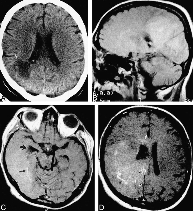
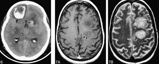
Background and purpose: Systemic invasive aspergillosis involves the brain through hematogenous dissemination. A retrospective review of 18 patients with aspergillosis involving the brain was performed in order to present imaging findings and thereby broaden the understanding of the distribution and imaging characteristics of brain Aspergillus infection and to facilitate its early diagnosis.

Methods: The neuroimaging studies of 17 biopsy- or autopsy-proved cases and one clinically diagnosed case were examined retrospectively by two neuroradiologists. The studies were evaluated for anatomic distribution of lesions, signal characteristics of lesions, enhancement, hemorrhage, and progression on serial studies (when performed). Medical records, biopsy reports, and autopsy findings were reviewed.

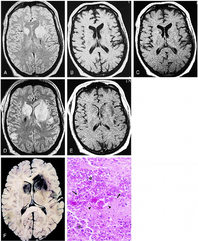
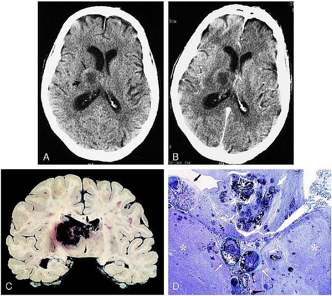
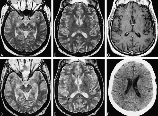
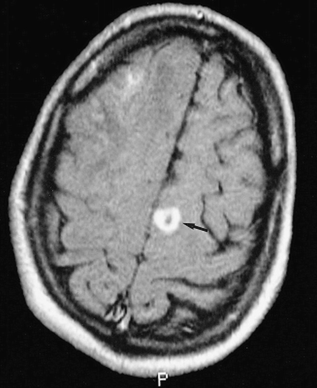
Results: Thirteen of 18 patients had involvement of the basal nuclei and/or thalami. Nine of the 10 patients with lesions at the corticomedullary junction also had lesions in the basal nuclei or thalami. Callosal lesions were seen in seven patients. Progression of lesion number and size was seen in all 11 patients in whom serial studies had been performed. Enhancement was minimal or absent in most cases. There was gross hemorrhage in eight of the 18, and definite ring-enhancement in three.

Conclusion: Among our cases, lesions in perforating artery territories were more common than those at the corticomedullary junction. Ring enhancement and gross hemorrhage may be present, but are not necessary for the prospective diagnosis.


Figures

<sc>fig</sc> 1.
fig 1.
48-year-old man, 3 weeks after liver transplantation, with decline in mental status and flaccidity in bilateral lower extremities and left upper extremity. A–C, Axial proton density–weighted (3000/25/2 [TR/TE/excitations]) (A) and unenhanced (500/20/2) (B) and contrast-enhanced (530/20/2) (C) T1-weighted spin-echo images. Areas of hyperintensity are present in the basal nuclei bilaterally, as well as in the left internal capsule on the proton density–weighted image (arrows, A), with corresponding modest hypointensity on the T1-weighted images. A small focus of enhancement is seen in the right caudate head (arrow, C). D and E, Axial proton density–weighted (3000/25/2) and contrast-enhanced T1-weighted (530/20/2) spin-echo images obtained 6 days later show new involvement of the right internal capsule (large solid arrow) and callosal genu (small solid arrow). Cortical and subcortical lesions are also now seen (open arrows). Although the lateral ventricles are now effaced, there is rather little enhancement. The patient died several hours after this study was obtained. F, The horizontally sectioned gross specimen shows red-brown areas of hemorrhagic necrosis corresponding to the abnormalities seen in D in the basal nuclei, and in the cortex and subcortical white matter. Hemorrhage is also seen in the genu of the corpus callosum. G, Histologic section shows extensive necrosis with infiltration by polymorphonuclear leukocytes and macrophages (asterisks). Numerous Aspergillus hyphae are scattered throughout the section (arrows), and vascular fibrinoid necrosis is present (arrowheads) (PAS, hematoxylin; original magnification ×40).
<sc>fig</sc> 2.
fig 2.
80-year-old man with chronic lymphocytic leukemia presented with disorientation and incontinence. A, Axial unenhanced CT scan shows a solitary lesion in the right thalamus (large arrow) with surrounding vasogenic edema. Attenuation is slightly increased in the anterolateral aspect of the lesion, indicating petechial hemorrhage (small arrow). Note central hypoattenuation. B, Corresponding axial contrast-enhanced CT scan shows no appreciable enhancement. The patient died 2 days later. C, Coronally sectioned brain shows bilateral hemorrhagic necrosis of the thalami (right greater than left) and fornices (arrow). Smaller hemorrhagic foci are present in the corpus callosum. D, Whole-mount coronal section shows thalamic necrosis bilaterally (asterisks). Hemorrhagic necrosis is present in the fornices (large white arrow). Thrombosis of the internal cerebral veins is present (small white arrows). A Gomori methenamine silver–stained section (not shown) revealed hyphae throughout the internal cerebral vein thrombus and walls and in the necrotic brain parenchyma. This anatomic distribution of lesions and the histologic findings indicate the primary pathophysiology is venous infarction with associated fungal encephalitis. Choroid plexitis is also seen (black arrow) (trichrome, original magnification ×1).
<sc>fig</sc> 3.
fig 3.
51-year-old man after bone marrow transplantation for IgA multiple myeloma with mental status decline. Pulmonary aspergillosis had been proved by bronchoalveolar lavage. A–C were obtained at neurologic presentation, D–F 5 months later. A, Axial fast spin-echo T2-weighted (2200/84eff /1) image shows a mesencephalic lesion centered in the left substantia nigra (arrow). B, Axial fast spin-echo T2-weighted (2200/84eff /1) image shows a hyperintense white matter lesion in the right parietal lobe (arrow). This patient also had severe (proved) bacterial sinusitis. C, Axial fast spin-echo T1-weighted (600/12/2) image with contrast shows no definite enhancement of the lesion (arrow). D, Axial fast spin-echo T2-weighted (2200/84eff /1) image of the midbrain shows return to normal after treatment with amphotericin B lipid complex. E, Axial fast spin-echo T2-weighted (2200/84eff /1) image shows marked central hypointensity at the site of the parietal white matter lesion (arrow). There was again no enhancement of this lesion (image not shown). F, Axial unenhanced CT scan shows high attenuation at the site of the parietal white matter lesion (arrow), confirming calcification of this lesion.
<sc>fig</sc> 4.
fig 4.
38-year-old man with seizures after a second liver transplantation. Axial contrast-enhanced spin-echo T1-weighted (630/20/2) image shows a ring-enhancing lesion at the corticomedullary junction of the left superior frontal gyrus (arrow). Biopsy of a corticomedullary junction lesion in the right frontal lobe (partially seen) confirmed cerebral aspergillosis
<sc>fig</sc> 5.
fig 5.
40-year-old woman after simultaneous pancreas/kidney transplantation had mental status decline and, later, a left homonymous hemianopia. A, Axial unenhanced CT scan shows a large low-attenuation lesion (arrow) extending to the right lateral ventricle. B, Parasagittal unenhanced spin-echo T1-weighted (630/20/2) MR image 10 days later shows a large, confluent area of diffuse, mild hyperintensity, suggesting petechial hemorrhage. C, Axial contrast-enhanced T1-weighted (450/20/2) MR image shows involvement of the temporal and occipital lobes. The right temporal horn is obstructed (large arrow). There are serpentine foci of enhancement (small arrow). D, Axial contrast-enhanced T1-weighted (450/20/2) MR image superior to C shows involvement of the frontal and parietal lobes, with focal areas of enhancement (black arrow). The abnormality extends into the corpus callosum (white arrow). The patient died 2 days later. At autopsy, this large abnormality was found to be Aspergillus cerebritis with scattered areas of petechial hemorrhage.
<sc>fig</sc> 6.
fig 6.
58-year-old woman after heart transplantation presented with obtundation and an unresponsive left pupil. Hemorrhagic lesions are seen on unenhanced CT scan in the left caudate head (small arrow) and right frontal lobe (large arrow). Aspergillosis was proved at autopsy. fig 7. 43-year-old man after bone marrow transplantation for acute myelogenous leukemia, presented with mental status decline. A, Axial fast spin-echo T1-weighted (600/12eff /2) MR image with contrast shows minimal punctate enhancement (arrows). There was no evidence of hemorrhage on an unenhanced T1-weighted MR image (not shown). B, The hemispheric white matter lesions are conspicuously hyperintense (arrows) with fast spin-echo T2-weighting (2200/84eff /1). The patient died 2 days later. At autopsy, these lesions consisted of hemorrhagic, necrotizing encephalitis with numerous branching hyphae.

Similar articles

Cited by

References

    1. Torre-Cisneros J, Lopez OL, Kusne S, et al. CNS aspergillosis in organ transplantation: a clinicopathological study. J Neurol Neurosurg Psychiatry 1993;56:188-193 - PMC - PubMed
    1. Pagano L, Ricci P, Montillo M, et al. Localization of aspergillosis to the central nervous system among patients with acute leukemia: report of 14 cases. Gruppo Italiano Malattie Ematologiche dell'Adulto Infection Program. Clin Infect Dis 1996;23:628-630 - PubMed
    1. Cox J, Murtagh FR, Wilfong A, Brenner J. Cerebral aspergillosis: MR imaging and histopathologic correlation. AJNR Am J Neuroradiol 1992;13:1489-1492 - PMC - PubMed
    1. Khoury H, Adkins D, Miller G, Goodnaugh L, Brown R, DiPerso J. Resolution of invasive central nervous system aspergillosis in a transplant recipient. Bone Marrow Transplant 1997;20:179-180 - PubMed
    1. Schwartz S, Milatovic D, Thiel E. Successful treatment of cerebral aspergillosis with a novel triazole (voriconazole) in a patient with acute leukemia. Br J Haematol 1997;97:663-665 - PubMed