

Incidental detection of hippocampal sclerosis on MR images: is it significant?
- PMID: 10543629
- PMCID: PMC7056185
Incidental detection of hippocampal sclerosis on MR images: is it significant?
Abstract
Background and purpose: The prevalence of hippocampal sclerosis in the general nonepileptic patient population is not well described. While reports of its association with partial complex seizures are abundant, its absence in nonafflicted patients is generally presumed but not well documented. To test the hypothesis that hippocampal sclerosis is specific for epilepsy, we reviewed the MR imaging studies of 207 patients referred for hearing loss to determine whether high-resolution MR imaging could detect unsuspected hippocampal sclerosis in nonepileptic patients.
Methods: Our institution screens patients with hearing loss by using high-resolution coronal and axial temporal bone MR imaging that includes the hippocampus within the imaging volume. We retrospectively reviewed 207 studies randomly selected from this database.
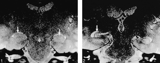
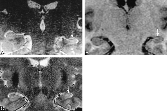
Results: The hippocampus was normal in 205 patients; in the remaining two patients we identified one or more primary determinants for hippocampal sclerosis. Subsequent retrospective chart review revealed that both patients had had previously diagnosed seizure disorders.
Conclusion: The imaging determinants of hippocampal sclerosis are not prevalent in nonepileptic patients. Incidental identification of hippocampal sclerosis on MR images is uncommon and significant, and should prompt further clinical investigation to exclude a seizure disorder.
Figures
Comment in
-
Incidental detection of hippocampal sclerosis.AJNR Am J Neuroradiol. 1999 Oct;20(9):1575-6. AJNR Am J Neuroradiol. 1999. PMID: 10543621 Free PMC article. No abstract available.
Similar articles
-
Anterior temporal changes on MR images of children with hippocampal sclerosis: an effect of seizures on the immature brain?AJNR Am J Neuroradiol. 2003 Sep;24(8):1670-7. AJNR Am J Neuroradiol. 2003. PMID: 13679290 Free PMC article.
-
Partial loss of hippocampal striation in medial temporal lobe epilepsy: pilot evaluation with high-spatial-resolution T2-weighted MR imaging at 3.0 T.Radiology. 2009 Jun;251(3):873-81. doi: 10.1148/radiol.2513080445. Epub 2009 Apr 3. Radiology. 2009. PMID: 19346512
-
Qualitative MR imaging of refractory temporal lobe epilepsy requiring surgery: correlation with pathology and seizure outcome after surgery.AJR Am J Roentgenol. 1997 Sep;169(3):875-82. doi: 10.2214/ajr.169.3.9275915. AJR Am J Roentgenol. 1997. PMID: 9275915
-
Epilepsy: the role of MR imaging.AJR Am J Roentgenol. 1992 Dec;159(6):1165-74. doi: 10.2214/ajr.159.6.1442376. AJR Am J Roentgenol. 1992. PMID: 1442376 Review.
-
Hippocampal pathology.Neuroimaging Clin N Am. 1997 Feb;7(1):51-65. Neuroimaging Clin N Am. 1997. PMID: 9100231 Review.
Cited by
-
New MRI Finding in Migraineurs: Mesial Temporal Sclerosis.J Biomed Phys Eng. 2020 Aug 1;10(4):459-466. doi: 10.31661/jbpe.v0i0.887. eCollection 2020 Aug. J Biomed Phys Eng. 2020. PMID: 32802794 Free PMC article.
-
Rare variants and de novo variants in mesial temporal lobe epilepsy with hippocampal sclerosis.Neurol Genet. 2018 Jun 11;4(3):e245. doi: 10.1212/NXG.0000000000000245. eCollection 2018 Jun. Neurol Genet. 2018. PMID: 29904720 Free PMC article.
-
Prevalence of hippocampal malrotation in a population without seizures.AJNR Am J Neuroradiol. 2009 Sep;30(8):1571-3. doi: 10.3174/ajnr.A1657. Epub 2009 Jun 18. AJNR Am J Neuroradiol. 2009. PMID: 19541778 Free PMC article.
-
Hippocampal abnormalities in an MR imaging series of patients with tuberous sclerosis.AJNR Am J Neuroradiol. 2010 Jun;31(6):1059-62. doi: 10.3174/ajnr.A1972. Epub 2010 Jan 6. AJNR Am J Neuroradiol. 2010. PMID: 20053803 Free PMC article.
-
Importance of genetic factors in the occurrence of epilepsy syndrome type: a twin study.Epilepsy Res. 2011 Nov;97(1-2):103-11. doi: 10.1016/j.eplepsyres.2011.07.018. Epub 2011 Aug 31. Epilepsy Res. 2011. PMID: 21885256 Free PMC article.
References
-
- Gloor P. The Temporal Lobe and Limbic System. . New York: Oxford University Press; 1997
-
- Kuzniecky R, Burgard S, Bilir E,, et al. Qualitative MRI segmentation in mesial temporal sclerosis: clinical correlations. . Epilepsia 1996;37:433-439 - PubMed
-
- Hayes C, Tsuruda J, Mathis C. Temporal lobes: surface MR coil phased-array imaging. . Radiology 1993;189:918-920 - PubMed
-
- Naidich T, Daniels D, Haughton V, Williams A, Pojunas K, Palacios E. Hippocampal formation and related structures of the limbic lobe, II: anatomic-MR correlation. . Radiology 1987;162:755-761 - PubMed
MeSH terms
LinkOut - more resources
Full Text Sources
Medical