

Inflammatory CNS demyelination: histopathologic correlation with in vivo quantitative proton MR spectroscopy
- PMID: 10543631
- PMCID: PMC7056180
Inflammatory CNS demyelination: histopathologic correlation with in vivo quantitative proton MR spectroscopy
Abstract
Background and purpose: The mechanisms behind the demyelination that is characteristic of multiple sclerosis (MS) are still poorly understood. The purpose of this study was to compare immunopathologic findings in demyelinating lesions of three patients with in vivo assessments obtained by quantitative proton MR spectroscopy (MRS).
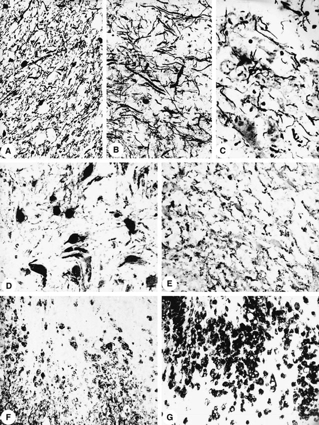
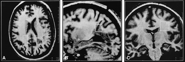
Methods: Between four and seven stereotactic needle brain biopsies were performed in three young adults with diagnostically equivocal findings for MS. Axonal density, gliosis, blood brain-barrier breakdown, and demyelinating activity of lesions were determined. Combined MR/MRS studies were performed (T1-weighted fast low-angle shot and single-voxel stimulated-echo acquisition mode), and absolute metabolite levels were obtained with a user-independent fitting routine. Metabolite control values were obtained from a group of age-matched healthy volunteers (n = 40, age range, 20-25 years old). Alterations of metabolite levels of control subjects were considered significant when exceeding two standard deviations.
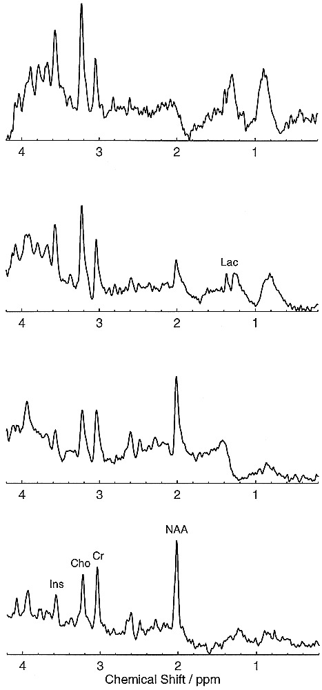
Results: There were parallel decreases of N-acetylaspartate (21%-82%) and reductions of axonal density (44%-74%) in demyelinating plaques. Concomitant increases of choline (75%-152%) and myo-inositol (84%-160%) corresponded to glial proliferation. Elevated lactate was associated with inflammation.
Conclusion: The present data suggest that in vivo MRS indicates key pathologic features of demyelinating lesions.
Figures
Similar articles
-
Axonal damage but no increased glial cell activity in the normal-appearing white matter of patients with clinically isolated syndromes suggestive of multiple sclerosis using high-field magnetic resonance spectroscopy.AJNR Am J Neuroradiol. 2007 Sep;28(8):1517-22. doi: 10.3174/ajnr.A0594. AJNR Am J Neuroradiol. 2007. PMID: 17846203 Free PMC article.
-
Axonal damage in multiple sclerosis plaques: a combined magnetic resonance imaging and 1H-magnetic resonance spectroscopy study.J Neurol Sci. 2001 Jan 1;182(2):143-50. doi: 10.1016/s0022-510x(00)00464-0. J Neurol Sci. 2001. PMID: 11137520
-
Quantitative proton magnetic resonance spectroscopy of focal brain lesions.Pediatr Neurol. 2000 Jul;23(1):22-31. doi: 10.1016/s0887-8994(00)00141-7. Pediatr Neurol. 2000. PMID: 10963966
-
The contribution of (1)H-magnetic resonance spectroscopy in defining the pathophysiology of multiple sclerosis.Ital J Neurol Sci. 1999;20(5 Suppl):S241-5. doi: 10.1007/s100729970004. Ital J Neurol Sci. 1999. PMID: 10662956 Review.
-
Proton MR spectroscopy in multiple sclerosis: value in establishing diagnosis, monitoring progression, and evaluating therapy.AJR Am J Roentgenol. 1991 Nov;157(5):1073-8. doi: 10.2214/ajr.157.5.1927795. AJR Am J Roentgenol. 1991. PMID: 1927795 Review.
Cited by
-
Brain Metabolite Changes in Patients with Relapsing-Remitting and Secondary Progressive Multiple Sclerosis: A Two-Year Follow-Up Study.PLoS One. 2016 Sep 16;11(9):e0162583. doi: 10.1371/journal.pone.0162583. eCollection 2016. PLoS One. 2016. PMID: 27636543 Free PMC article.
-
In vivo characterisation of soft tissue tumours by 1.5-T proton MR spectroscopy.Eur Radiol. 2012 May;22(5):1131-9. doi: 10.1007/s00330-011-2350-9. Epub 2011 Dec 4. Eur Radiol. 2012. PMID: 22138734
-
The role of nonconventional magnetic resonance imaging techniques in demyelinating disorders.Curr Neurol Neurosci Rep. 2003 May;3(3):238-45. doi: 10.1007/s11910-003-0084-z. Curr Neurol Neurosci Rep. 2003. PMID: 12691629 Review.
-
Evidence of widespread metabolite abnormalities in Myalgic encephalomyelitis/chronic fatigue syndrome: assessment with whole-brain magnetic resonance spectroscopy.Brain Imaging Behav. 2020 Apr;14(2):562-572. doi: 10.1007/s11682-018-0029-4. Brain Imaging Behav. 2020. PMID: 30617782 Free PMC article.
-
Dietary inulin alters the gut microbiome, enhances systemic metabolism and reduces neuroinflammation in an APOE4 mouse model.PLoS One. 2019 Aug 28;14(8):e0221828. doi: 10.1371/journal.pone.0221828. eCollection 2019. PLoS One. 2019. PMID: 31461505 Free PMC article.
References
-
- Lassmann H. Comparative Neuropathology of Chronic Experimental Allergic Encephalomyelitis and Multiple Sclerosis. Berlin, Heidelberg: Springer-Verlag; 1983 - PubMed
-
- Prineas J. W. The neuropathology of multiple sclerosis. In: Koetsier, J. C. Demyelinating Diseases Amsterdam: Elsevier Science Publishers; 1985;8:213-257
-
- McDonald WI, Miller DH, Barnes D. The pathological evolution of multiple sclerosis. Neuropathol Appl Neurobiol 1992;18:319-334 - PubMed
-
- Miller DH, Grossman RI, Reingold SC, McFarland HF. The role of magnetic resonance techniques in understanding and managing multiple sclerosis. Brain 1998;121:3-24 - PubMed
Publication types
MeSH terms
Substances
LinkOut - more resources
Full Text Sources
Medical