

Case Reports
Clinically aggressive diffuse capillary telangiectasia of the brain stem: a clinical radiologic-pathologic case study
Affiliations
- PMID: 10543639
- PMCID: PMC7056202
Item in Clipboard
Case Reports
Clinically aggressive diffuse capillary telangiectasia of the brain stem: a clinical radiologic-pathologic case study
AJNR Am J Neuroradiol.
1999 Oct.
Abstract
Capillary malformations or telangiectasias of the brain usually exhibit a benign clinical course, although occassionally they may be associated with mild to moderate symptomatology of uncertain origin. We report a case of an exceptionally aggressive capillary telangiectasia in a child, which was associated with progressive neurologic deterioration resulting in death.
Figures
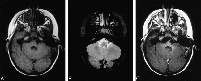
A, Axial T1-weighted (700/17/1) MR image shows mild asymmetry of the lower basis pontis and middle cerebellar peduncle on the left. Note the geographic region of subtle hypointensity centrally and to the left of midline and questionable punctate hyperintense foci near the midline. B, Axial T2-weighted (2200/90/1) image showing abnormal hyperintensity within the left ventral portion of the brain stem at the level of the inferior cerebellar peduncle/medulla. This focus is slightly caudal to the region of hypointensity seen in figure 1B. C, Post-gadolinium T1-weighted (700/19/1) axial image reveals homogeneous enhancement within the rostral pons and adjacent cerebellar peduncles. The magnitude of contrast enhancement is disproportionately greater than the focal signal changes seen on the T2-weighted images (see figure 1B).
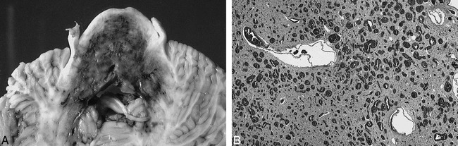
A, Gross anatomic specimen showing axial section through mid-pontine level. The capillary malformation is clearly demarcated by the extensive bluish discoloration involving nearly the entire cross-sectional area of the basis pons and tegmentum with extension into the middle cerebellar peduncles. There are patchy areas of relatively more dense discoloration representing increased density of telangiectasia. B, Microscopic specimen showing the basis pontis. Vast numbers of blood-filled endothelium-lined vascular channels measuring 50–150 microns in diameter have replaced much of the brain parenchyma. There is also gliosis and neuronal drop-out, but no evidence of hemorrhage.
Similar articles
-
Symptomatic capillary telangiectasia of the pons and intracerebral developmental venous anomaly - A rare association.Coll Antropol. 2011 Jan;35 Suppl 1:333-8. Coll Antropol. 2011. PMID: 21648357
-
Presumed capillary telangiectasia of the pons: MRI and follow-up.Eur Radiol. 2000;10(6):945-50. doi: 10.1007/s003300051043. Eur Radiol. 2000. PMID: 10879709
-
Capillary telangiectasias: clinical, radiographic, and histopathological features. Clinical article.J Neurosurg. 2010 Oct;113(4):709-14. doi: 10.3171/2009.9.JNS09282. J Neurosurg. 2010. PMID: 19817536
-
[Capillary telangiectasia].Radiologie (Heidelb). 2022 Aug;62(8):654-658. doi: 10.1007/s00117-022-01037-z. Epub 2022 Jul 6. Radiologie (Heidelb). 2022. PMID: 35792920 Review. German.
-
Symptomatic unruptured capillary telangiectasia of the brain stem: report of three cases and review of the literature.J Neurol Neurosurg Psychiatry. 2001 Sep;71(3):390-3. doi: 10.1136/jnnp.71.3.390. J Neurol Neurosurg Psychiatry. 2001. PMID: 11511717 Free PMC article. Review.
Cited by
-
Pontine capillary telangiectasia as visualized on MR imaging causing a clinical picture resembling basilar-type migraine: a case report.J Neurol. 2009 Oct;256(10):1775-7. doi: 10.1007/s00415-009-5204-5. Epub 2009 Jun 17. J Neurol. 2009. PMID: 19533201 Free PMC article.
-
A Rare Association of Trigeminal Autonomic Cephalgia: Pontine Capillary Telangiectasia.Neuroradiol J. 2015 Apr;28(2):145-7. doi: 10.1177/1971400915576656. Epub 2015 May 11. Neuroradiol J. 2015. PMID: 25963152 Free PMC article.
-
Isolated developmental venous anomaly of the pons with transpontine drainage: case report.Clin Neuroradiol. 2014 Mar;24(1):77-81. doi: 10.1007/s00062-013-0206-1. Epub 2013 Feb 9. Clin Neuroradiol. 2014. PMID: 23397208 No abstract available.
-
Aggressive giant pontine capillary telangiectasia with hypertrophic olivary degeneration and brainstem atrophy: illustrative case.J Neurosurg Case Lessons. 2025 Mar 24;9(12):CASE24746. doi: 10.3171/CASE24746. Print 2025 Mar 24. J Neurosurg Case Lessons. 2025. PMID: 40127484 Free PMC article.
-
Cerebral capillary telangiectasias: a meta-analysis and review of the literature.Neurosurg Rev. 2013 Apr;36(2):187-93; discussion 194. doi: 10.1007/s10143-012-0435-9. Epub 2012 Nov 29. Neurosurg Rev. 2013. PMID: 23192650 Review.
References
-
- Okazaki H. Cerebrovascular Disease. In: Fundamentals of Neuropathology-Morphologic Basis of Neurologic Disorders. 2nd ed. New York: IGAKU-SHOIN Medical Publishers; 1989;27-94
-
- Chang SD, Steinberg GK, Rosario M, Crowley RS, Hevner RF. Mixed arteriovenous malformation and capillary telangectasia: a rare subset of mixed vascular malformations (case report). J Neurosurg 1997;86:699-703 - PubMed
-
- Lee RR, Becher MW, Benson ML, Rigamonti D. Brain Capillary Telangiectasia: MR Imaging Apearance and Clinicohistopathologic Findings. Radiology 1997;205:797-805 - PubMed
-
- Rigamonti D, Johnson PC, Spetzler RF, Hadley MN, Drayer BP. Cavernous Malformations and Capillary Telangiectasia: a spectrum within a single pathological entity. Neurosurgery 1991;28:60-64 - PubMed
Publication types
MeSH terms
LinkOut - more resources
Full Text Sources
Medical