

Neurointerventional experience with an arteriotomy suture device
Affiliations
- PMID: 10543645
- PMCID: PMC7056203
Item in Clipboard
Neurointerventional experience with an arteriotomy suture device
AJNR Am J Neuroradiol.
1999 Oct.
Abstract
We describe our experience with an arteriotomy closure device that has become a routine tool for the management of most patients in our neurointerventional service. In our experience, this device contributes significantly to patient comfort by allowing mobilization within 2 hours of a procedure, even with anticoagulants. Efficacy and safety of this suture device requires proctoring during initial experience.
Figures
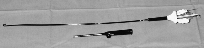
The device resembles a J-shaped 40-cm sheath with hydrophilic coating. It is inserted over a wire into the artery. Package also includes a knot pusher used for achieving tight closure of sutures under subcutaneous tissues

Hub of the device features a translucent ring handle for retracting the needles out of the shaft. A clear marker tubing (white arrow) emits pulsatile flow of blood when device is fully in position within vessel. Prior to deployment of the needles, slack from sutures is housed within two other clear tubings (black arrowheads) extending from the hub

After anesthetization of groin area with lidocaine and epinephrine, the device is dissected through the subcutaneous tissues until the narrow shaft is fully within the lumen of the artery. A hole near end of shaft allows escape of arterial flow into the clear tubing extending from hub (white arrow in 2), to indicate to the operator that the device is in place

A and B, Needles are retracted from shaft by pulling on ring handle. As they emerge from shaft (close-up view A), needles bevel away from shaft so as to pierce edges of the arteriotomy site, and then enter the hub. Then they emerge from hub with suture strings attached (black arrowhead in B). A forceps is used to withdraw needles and sutures from hub completely. Note that thread is attached to the end of needle, which emerges first from the hub (arrow)

A and B, needles are cut from sutures and the device is withdrawn partially to give the operator control of sutures on the arterial side of hub. An improved clinch knot (A) or a series of surgeon's knots are applied loosely and tightened against the arterial wall as the device is withdrawn from artery (B). The knot pusher can be used then to tighten the knot
Similar articles
-
Safety and efficacy of a 6 French perclose arterial suturing device following percutaneous coronary interventions: a pilot evaluation.J Invasive Cardiol. 2002 Dec;14(12):741-5. J Invasive Cardiol. 2002. PMID: 12454337
-
Initial experience using Prostar: a new device for percutaneous suture-mediated closure of arterial puncture sites.Cathet Cardiovasc Diagn. 1996 Apr;37(4):367-72. doi: 10.1002/(SICI)1097-0304(199604)37:4<367::AID-CCD5>3.0.CO;2-9. Cathet Cardiovasc Diagn. 1996. PMID: 8721692
-
[First experience with the use of a clip device for closure of arterial approach after endovascular treatment of coronary arteries].Kardiologiia. 2008;48(1):15-8. Kardiologiia. 2008. PMID: 18260990 Russian.
-
Current developments in percutaneous arterial closure devices.Ann Vasc Surg. 2000 Nov;14(6):683-7. doi: 10.1007/s100169910123. Ann Vasc Surg. 2000. PMID: 11128469 Review. No abstract available.
-
Angioplasty and stenting in the carotid and vertebral arteries.Postgrad Med J. 1998 Jan;74(867):7-10. doi: 10.1136/pgmj.74.867.7. Postgrad Med J. 1998. PMID: 9538479 Free PMC article. Review.
Cited by
-
The safety and efficacy of the Angio-Seal closure device in diagnostic and interventional neuroangiography setting: a single-center experience with 1,443 closures.Neuroradiology. 2007 Sep;49(9):739-46. doi: 10.1007/s00234-007-0249-6. Epub 2007 Jun 27. Neuroradiology. 2007. PMID: 17594084
-
Prospective comparison of angio-seal versus manual compression for hemostasis after neurointerventional procedures under systemic heparinization.AJNR Am J Neuroradiol. 2013 Feb;34(2):397-401. doi: 10.3174/ajnr.A3226. Epub 2012 Aug 2. AJNR Am J Neuroradiol. 2013. PMID: 22859279 Free PMC article. Clinical Trial.
-
An evaluation of immediate sheath removal and use of the Angio-Seal vascular closure device in neuroradiological interventions.Neuroradiology. 2006 Jan;48(1):45-9. doi: 10.1007/s00234-005-0013-8. Epub 2005 Oct 28. Neuroradiology. 2006. PMID: 16261336
-
Transcervical access via direct neck exposure for neurointerventional procedures in the hybrid angiosuite.Neuroradiology. 2018 May;60(5):565-573. doi: 10.1007/s00234-018-1994-4. Epub 2018 Mar 1. Neuroradiology. 2018. PMID: 29497785
References
-
- Dion JE, Gates PC, Fox AJ, et al. Clinical events following neuroangiography: a prospective study. Stroke 1987;18:997-1004 - PubMed
-
- Olivecrona H. Complications of cerebral angiography. Neuroradiology 1977;14:175-181 - PubMed
-
- Uchino A. Local complication in transbrachial cerebral angiography using the 4-F catheter. Neurolo Med Chir (Tokyo) 1991;31:647-649 - PubMed
-
- Millward SF. Letter to the editor: Routine transbrachial angiography. Radiology 1989;172:577 - PubMed
-
- Fruhwirth J, Pascher O, Haufer H, Amann W. Local vascular complications after iatrogenic femoral artery puncture. Wiener Klinische Wochenschrift 1996;108:196-200 - PubMed
MeSH terms
LinkOut - more resources
Full Text Sources