

Epitope identification for a panel of anti-Sinorhizobium meliloti monoclonal antibodies and application to the analysis of K antigens and lipopolysaccharides from bacteroids
- PMID: 10543844
- PMCID: PMC91702
- DOI: 10.1128/AEM.65.11.5186-5191.1999
Epitope identification for a panel of anti-Sinorhizobium meliloti monoclonal antibodies and application to the analysis of K antigens and lipopolysaccharides from bacteroids
Abstract
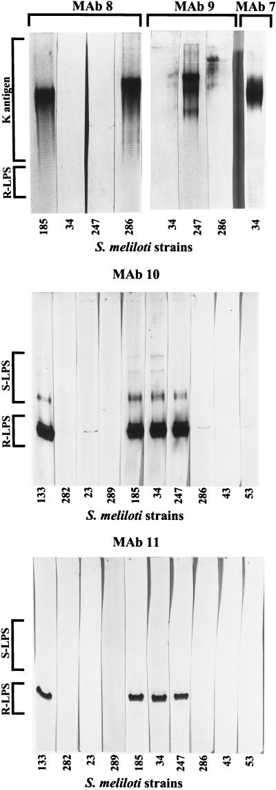
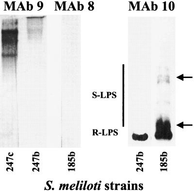
In two published reports using monoclonal antibodies (MAbs) generated against whole cells, Olsen et al. showed that strain-specific antigens on the surface of cultured cells of Sinorhizobium meliloti were diminished or absent in the endophytic cells (bacteroids) recovered from alfalfa nodules, whereas two common antigens were not affected by bacterial differentiation (P. Olsen, M. Collins, and W. Rice, Can. J. Microbiol. 38:506-509, 1992; P. Olsen, S. Wright, M. Collins, and W. Rice, Appl. Environ. Microbiol. 60:654-661, 1994). The nature of the antigens (i.e., the MAb epitopes), however, were not determined in those studies. For this report, the epitopes for five of the anti-S. meliloti MAbs were identified by polyacrylamide gel electrophoresis-immunoblot analyses of the polysaccharides extracted from S. meliloti and Sinorhizobium fredii. This showed that the strain-specific MAbs recognized K antigens, whereas the strain-cross-reactive MAbs recognized the lipopolysaccharide (LPS) core. The MAbs were then used in the analysis of the LPS and K antigens extracted from S. meliloti bacteroids, which had been recovered from the root nodules of alfalfa, and the results supported the findings of Olsen et al. The size range of the K antigens from bacteroids of S. meliloti NRG247 on polyacrylamide gels was altered, and the epitope was greatly diminished in abundance compared to those from the cultured cells, and no K antigens were detected in the S. meliloti NRG185 bacteroid extract. In contrast to the K antigens, the LPS core appeared to be similar in both cultured cells and bacteroids, although a higher proportion of the LPS fractionated into the organic phase during the phenol-water extraction of the bacteroid polysaccharides. Importantly, immunoblot analysis with an anti-LPS MAb showed that smooth LPS production was modified in the bacteroids.
Figures
Similar articles
-
Sinorhizobium fredii and Sinorhizobium meliloti produce structurally conserved lipopolysaccharides and strain-specific K antigens.Appl Environ Microbiol. 1998 Dec;64(12):4930-8. doi: 10.1128/AEM.64.12.4930-4938.1998. Appl Environ Microbiol. 1998. PMID: 9835585 Free PMC article.
-
The Lipid A substructure of the Sinorhizobium meliloti lipopolysaccharides is sufficient to suppress the oxidative burst in host plants.New Phytol. 2005 Feb;165(2):559-65. doi: 10.1111/j.1469-8137.2004.01214.x. New Phytol. 2005. PMID: 15720666
-
Sinorhizobium fredii HH103 bacteroids are not terminally differentiated and show altered O-antigen in nodules of the Inverted Repeat-Lacking Clade legume Glycyrrhiza uralensis.Environ Microbiol. 2016 Sep;18(8):2392-404. doi: 10.1111/1462-2920.13101. Epub 2015 Dec 21. Environ Microbiol. 2016. PMID: 26521863
-
Structure of the unusual Sinorhizobium fredii HH103 lipopolysaccharide and its role in symbiosis.J Biol Chem. 2020 Aug 7;295(32):10969-10987. doi: 10.1074/jbc.RA120.013393. Epub 2020 Jun 16. J Biol Chem. 2020. PMID: 32546484 Free PMC article.
-
[Root Nodule Bacteria Sinorhizobium meliloti: Tolerance to Salinity and Bacterial Genetic Determinants].Mikrobiologiia. 2015 May-Jun;84(3):263-80. Mikrobiologiia. 2015. PMID: 26263687 Review. Russian.
Cited by
-
Analysis of the chromosome sequence of the legume symbiont Sinorhizobium meliloti strain 1021.Proc Natl Acad Sci U S A. 2001 Aug 14;98(17):9877-82. doi: 10.1073/pnas.161294398. Epub 2001 Jul 31. Proc Natl Acad Sci U S A. 2001. PMID: 11481430 Free PMC article.
-
Strain-ecotype specificity in Sinorhizobium meliloti-Medicago truncatula symbiosis is correlated to succinoglycan oligosaccharide structure.J Bacteriol. 2007 Nov;189(21):7733-40. doi: 10.1128/JB.00739-07. Epub 2007 Aug 31. J Bacteriol. 2007. PMID: 17766412 Free PMC article.
-
Lipopolysaccharides in diazotrophic bacteria.Front Cell Infect Microbiol. 2014 Sep 3;4:119. doi: 10.3389/fcimb.2014.00119. eCollection 2014. Front Cell Infect Microbiol. 2014. PMID: 25232535 Free PMC article. Review.
-
Structural characterization of a flavonoid-inducible Pseudomonas aeruginosa A-band-like O antigen of Rhizobium sp. strain NGR234, required for the formation of nitrogen-fixing nodules.J Bacteriol. 2005 Sep;187(18):6479-87. doi: 10.1128/JB.187.18.6479-6487.2005. J Bacteriol. 2005. PMID: 16159781 Free PMC article.
-
Flavonoid-inducible modifications to rhamnan O antigens are necessary for Rhizobium sp. strain NGR234-legume symbioses.J Bacteriol. 2006 May;188(10):3654-63. doi: 10.1128/JB.188.10.3654-3663.2006. J Bacteriol. 2006. PMID: 16672619 Free PMC article.
References
-
- Becquart-de Kozak I, Reuhs B L, Buffard D, Breda C, Kim J S, Esnault R, Kondorosi A. Role of the K-antigen subgroup of capsular polysaccharides in the early recognition process between Rhizobium meliloti and alfalfa leaves. Mol Plant-Microbe Interact. 1997;10:114–123.
-
- Carlson R W, Reuhs B L, Forsberg L S, Kannenberg E L. Rhizobial cell surface carbohydrates. In: Goldberg J B, editor. Genetics of bacterial polysaccharides. Boca Raton, Fla: CRC Press; 1999. pp. 53–90.
-
- Corzo J, Pérez-Galdona R, León-Barrios M, Gutiérrez-Navarro A M. Alcian Blue fixation allows silver staining of the isolated polysaccharide component of bacterial lipopolysaccharides in polyacrylamide gels. Electrophoresis. 1991;12:439–441. - PubMed
-
- Denarie J, Roche P. Rhizobium nodulation signals. In: Verma D P S, editor. Molecular signals in plant-microbe communications. Boca Raton, Fla: CRC Press; 1992. pp. 295–324.
Publication types
MeSH terms
Substances
LinkOut - more resources
Full Text Sources
