. 1999 Nov 1;520 Pt 3(Pt 3):737-44.
doi: 10.1111/j.1469-7793.1999.00737.x.

Action of internal pronase on the f-channel kinetics in the rabbit SA node

Affiliations

Action of internal pronase on the f-channel kinetics in the rabbit SA node

A Barbuti et al. J Physiol. .

Abstract

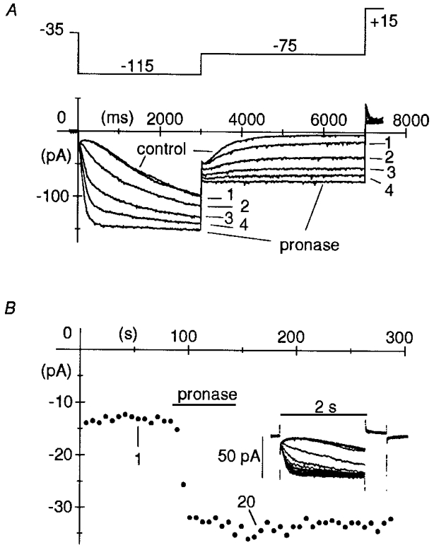
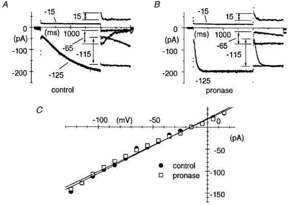
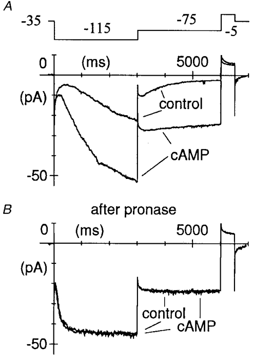
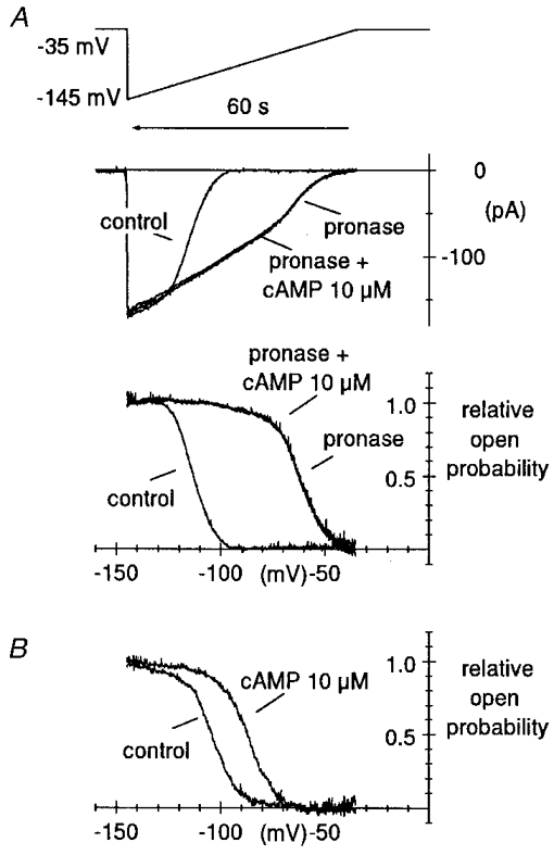
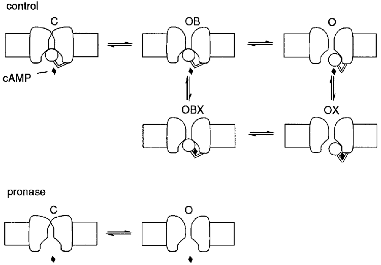
1. The hyperpolarization-activated If current was recorded in inside-out macropatches from sino-atrial (SA) node myocytes during exposure of their intracellular side to pronase, in an attempt to verify if cytoplasmic f-channel domains are involved in both voltage- and cAMP-dependent gating. 2. Superfusion with pronase caused a quick, dramatic acceleration of channel opening upon hyperpolarization and slowing, rapidly progressing into full blockade, of channel closing upon depolarization; these changes persisted after wash off of pronase and were irreversible, indicating proteolytic cleavage of channel regions which contribute to gating. 3. If recorded from patches normally responding to cAMP became totally insensitive to cAMP following pronase treatment, indicating partial or total removal of channel regions involved in the cAMP-dependent activation. 4. The fully activated I-V relationship was not modified by pronase, indicating that internal proteolysis did not affect the f-channel conductance. 5. The changes in If kinetics induced by pronase were due to a large depolarizing shift of the f-channel open probability curve (56.5 +/- 1.1 mV, n = 7). 6. These results are consistent with the hypothesis that cytoplasmic f-channel regions are implicated in dual voltage- and cAMP-dependent gating; also, since pronase does not abolish hyperpolarization-activated opening, an intrinsic voltage-dependent gating mechanism must exist which is inaccessible to proteolytic cleavage. A model scheme able to account for these data thus includes an intrinsic gating mechanism operating at depolarized voltages, and a blocking mechanism coupled to cAMP binding to the channel.


Figures

Figure 1
Figure 1. Action of pronase on macropatch If
A, pronase (2 mg ml−1) was superfused on the intracellular patch side for 1 min, after which superfusion with normal intracellular-like solution was resumed, while activating/ deactivating If current traces were recorded during two-step protocols (see top panel illustrating the voltage protocol) to −115 mV (3 s) and −75 mV (4 s) applied every 10 s. Holding potential was −35 mV. A short 0.5 s step to +15 mV was applied after each pair of hyperpolarizations to fully deactivate If. Pronase induced progressive acceleration of activation at −115 mV, and slowing of deactivation at −75 mV, until after about 60 s, deactivation was fully blocked. Plotted are the first four records following switch-on of pronase superfusion and the records in control and at steady state after pronase, as indicated. B, time course of pronase action. Pronase was superfused for 1 min while applying 2 s steps to −95 mV to activate If (followed by 0.5 s steps to +15 mV to achieve full deactivation prior to subsequent activating step) every 6 s, from a holding potential of −25 mV. In the inset, 20 successive records are superimposed showing the progressive acceleration of current activation. The plot represents If amplitude at −95 mV. The amplitudes of 1st and 20th record of inset are indicated.
Figure 4
Figure 4. Lack of modification of the If conductance by pronase
A and B, the fully activated I–V relationship of If was measured in control conditions (A) and after pronase treatment (B) according to a method previously developed (DiFrancesco et al. 1986), which consisted of two-step protocols where the current was either fully activated (by stepping to −125 mV for 2 s) or fully deactivated (by stepping to −15 mV for 2 s) before stepping to test potentials in the range −115 to +15 mV. In A and B, pairs of records shown correspond to test potentials of −115, −65 and +15 mV as indicated. Notice acceleration of current activation and slowing or block of current deactivation after pronase. C, fully activated I–V relationships in control conditions (•) and after pronase (□). The points were measured as differences between initial current values of pairs of traces corresponding to the same test potential (arrows in A and B). Linear regressions (straight lines) yielded reversal potentials of −15.7 and −15.1 mV, and conductances of 1.25 and 1.20 nS for control and pronase data, respectively.
Figure 2
Figure 2. Lack of action of cAMP on pronase-modified If
Macropatch If was recorded during two-step protocols (as shown in the top panel in A) from a holding potential of −35 mV to −115 and to −75 mV before (A) and after pronase (B). If responded normally to cAMP (10 μM) in control conditions (A); following pronase treatment, however, If activation was accelerated and deactivation blocked, and the current became completely insensitive to cAMP (B).
Figure 3
Figure 3. Large depolarizing shift of f-channel open probability curve induced by pronase
A, curves of the relative channel open probability (Po), normalized to maximum channel open probability, were measured by a voltage-ramp method previously developed (DiFrancesco & Mangoni, 1994) in control condition and after pronase treatment, as indicated. Ramps were applied from a holding potential of −35 mV to −145 mV and lasted 60 s (top panel). Notice that time runs backward for comparison with the open probability curves in the bottom panel. Ramp currents recorded in control conditions and after pronase, the latter with and without cAMP, are shown in the middle panel. Fitting open probability curves by the second power of the Boltzmann equation (see Methods) yielded V½ = −107.7 mV, v = 5.48 mV and V½ = −55.2 mV, v = 8.02 mV for the control and pronase curve, respectively (shift = 52.5 mV). The Po curve was not modified by 10 μM cAMP after pronase treatment. B, the same protocol was applied to determine, for comparison, the action of cAMP in a pronase-untreated macropatch. Best fitting parameters were V½ = −96.8 mV, v = 8.44 mV and V½ = −79.9 mV, v = 8.61 mV for the control and cAMP curve, respectively (shift = 16.9 mV).
Figure 5
Figure 5. Model interpretation of the action of pronase on f-channel gating
Upper panel (control), a terminal ball region is assumed to switch on voltage hyperpolarization from an open-blocked (OB) configuration, where it blocks the internal channel mouth, to an open one (O), the latter binding one cAMP molecule (OX) with higher affinity than the former (OBX). cAMP binding locks the ball in the open state. Symbols OB, O, OBX and OX refer to the allosteric model of DiFrancesco (, Fig. 2; replacing O for R, relaxed and OB for T, tense). Lower panel (pronase), protease cleavage of the terminal ball region abolishes the corresponding gating mechanism and the cAMP dependence. A second, voltage-dependent gating mechanism inaccessible to pronase is assumed to underly voltage-dependent kinetics between states C (closed) and O in the absence of the ‘ball’-dependent gating. Further explanation in text.

References

    1. Accili EA, DiFrancesco D. Inhibition of the hyperpolarization-activated current (if) of rabbit SA node myocytes by niflumic acid. Pflügers Archiv. 1996;431:757–762. - PubMed
    1. Baukrowitz T, Yellen G. Modulation of K+ current by frequency and external [K+]: a tale of two inactivation mechanisms. Neuron. 1995;15:951–960. - PubMed
    1. Clapham D. Not so funny anymore: pacing channels are cloned. Neuron. 1998;21:5–7. - PubMed
    1. Covarrubias M, Wei A, Salkoff L. Shaker, Shal, Shab and Shaw express independent K+ current systems. Neuron. 1991;7:763–773. - PubMed
    1. DiFrancesco D. Pacemaker mechanisms in cardiac tissue. Annual Reviews of Physiology. 1993;55:451–467. - PubMed

Publication types

Grants and funding

LinkOut - more resources