. 1999 Nov 1;520 Pt 3(Pt 3):761-9.
doi: 10.1111/j.1469-7793.1999.00761.x.

Subatmospheric pressure in the rabbit pleural lymphatic network

Affiliations

Subatmospheric pressure in the rabbit pleural lymphatic network

D Negrini et al. J Physiol. .

Abstract

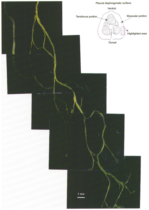
1. Hydraulic pressure in intercostal and diaphragmatic lymphatic vessels was measured through the micropuncture technique in 23 anaesthetised paralysed rabbits. Pleural lymphatic vessels with diameters ranging from 55 to 950 microm were observed under stereomicroscope view about 3-4 h after intrapleural injection of 20 % fluorescent dextrans. 2. Lymphatic pressure oscillated from a minimum (Pmin) to a maximum (Pmax) value, reflecting oscillations in phase with cardiac activity (cardiogenic oscillations) and lymphatic myogenic activity. With intact pleural space, Pmin in submesothelial diaphragmatic lymphatic vessels of the lateral apposition zone was -9.1 +/- 4.2 mmHg, more subatmospheric than the simultaneously recorded pleural liquid pressure amounting to -3.9 +/- 1.2 mmHg. In extrapleural intercostal lymphatic vessels Pmin averaged -1.3 +/- 2. 7 mmHg. 3. Cardiogenic pressure oscillations (Pmax - Pmin), were observed in all recordings; their mean amplitude was about 5 mmHg and was not dependent upon frequency of cardiac contraction, nor lymphatic vessel diameter, nor the Pmin value. 4. Intrinsic contractions of lymphatic vessel walls caused spontaneous pressure waves of about 7 mmHg in amplitude at a rate of 8 cycles min-1. 5. These results demonstrated the ability of pleural lymphatic vessels to generate pressure oscillations driving fluid from the subatmospheric pleural space into the lymphatic network.


Figures

Figure 1
Figure 1. Microphotograph of a loop of confluent lymphatic vessels on the pleural surface of the diaphragm, visualised through a stereomicroscope at about 4 h after intrapleural injection of fluorescent dextrans
Unidirectional valves separating adjacent lymphangions are clearly detectable. The tip of the recording micropipette can be seen in the central bottom part of the microphotograph. Scale bar corresponds to 50 μm.
Figure 2
Figure 2
Microphotographic reconstruction of the lymphatic diaphragmatic network on the lateral right side of the pleural diaphragm (see schematic drawing at the top right).
Figure 3
Figure 3. Simultaneous tracings of hydraulic pressures obtained with intact pleural space
From the top: central venous pressure, systemic arterial pressure, pleural liquid pressure (Pliq) in the costal apposition zone, pleural lymphatic pressure (Plymph) recorded in a diaphragmatic lymphatic vessel of 100 μm diameter (all values in mmHg).
Figure 4
Figure 4. Plymph tracers from two diaphragmatic lymphatic vessels with diameter of 300 μm (top panel) and 150 μm (bottom panel), respectively
Top panel: the observed pressure oscillations (cardiogenic oscillations) are in phase with cardiac stroke, Plymph shifting from a minimum (Pmin) to a maximum (Pmax) value. Bottom panel: slower pressure waves due to spontaneous myogenic contraction of the lymphatic vessel walls are superimposed onto the cardiogenic oscillations.
Figure 5
Figure 5. Effect of cardiogenic oscillations on Plymph
Intercostal (^, n = 94), diaphragmatic with intact pleural space (▴, n = 7) and diaphragmatic with open pleural space (▪, n = 56) Pmax values are plotted as a function of the corresponding Pmin. Continuous and dashed lines correspond, respectively, to the linear correlation through the pooled data and to the 95 % confidence limit of the correlation.
Figure 6
Figure 6
Bottom panel: schematic drawing of the functional arrangement of the pleural lymphatic vessels. Pleural fluid is drained via the stomas into the submesothelial lacunae of the initial lymphatic network. Initial lymphatic vessels empty into a net of extrapleural lymphatic vessels progressively more distant from the pleural space. Top panel: hydraulic pressures measured in the compartments presented in the bottom panel. ♦, pleural liquid pressure in the costal apposition zone. ▴, diaphragmatic Pmin and ▵, diaphragmatic Pmax with intact pleural space. •, intercostal Pmin and ^, intercostal Pmax.formula image, central venous pressure. Bars represent ± 1 s.d.

References

    1. Agostoni E. Mechanics of the pleural space. Physiological Reviews. 1972;52:57–128. - PubMed
    1. Benoit JN, Zawieja DC, Goodman AH, Granger HJ. Characterization of intact mesenteric lymphatic pump and its responsiveness to acute edemagenic stress. American Journal of Physiology. 1989;257:H2059–2069. - PubMed
    1. Clough G, Smaje LH. Simultaneous measurements of pressure in the interstitium and the terminal lymphatics of the cat mesentery. The Journal of Physiology. 1978;283:457–468. - PMC - PubMed
    1. Crowe MJ, von der Weid P-Y, Brock JA, van Helden DF. Co-ordination of contractile activity in guinea-pig mesenteric lymphatics. The Journal of Physiology. 1997;500:235–244. - PMC - PubMed
    1. Lee JS. Tissue fluid pressure, lymph pressure and fluid transport in rat intestinal villi. Microvascular Research. 1986;31:170–183. - PubMed

Publication types

LinkOut - more resources