. 1999 Nov 1;147(3):545-58.
doi: 10.1083/jcb.147.3.545.

Sphingosine kinase expression increases intracellular sphingosine-1-phosphate and promotes cell growth and survival

Affiliations

Sphingosine kinase expression increases intracellular sphingosine-1-phosphate and promotes cell growth and survival

A Olivera et al. J Cell Biol. .

Abstract

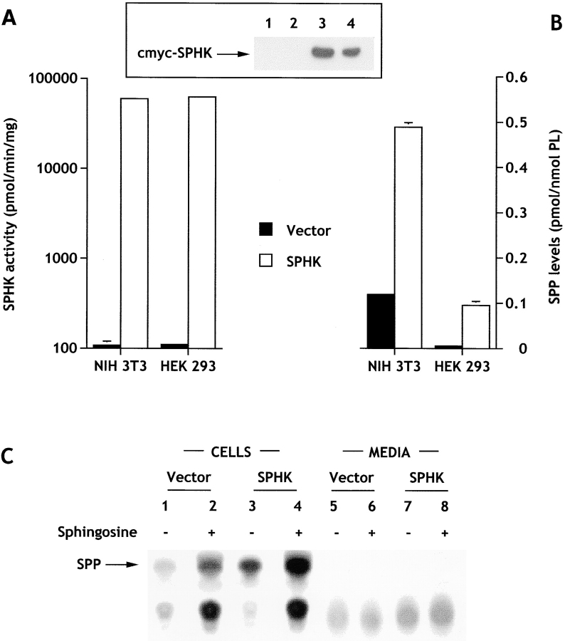
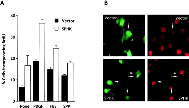
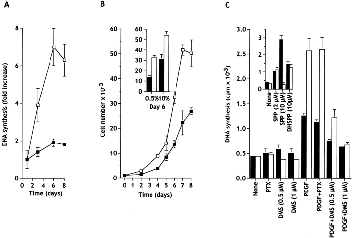
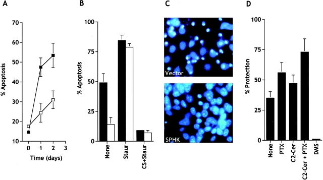
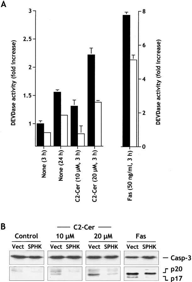
Sphingosine-1-phosphate (SPP) is a bioactive lipid that has recently been identified as the ligand for the EDG family of G protein-coupled cell surface receptors. However, the mitogenic and survival effects of exogenous SPP may not correlate with binding to cell-surface receptors (Van Brocklyn, J.R., M.J. Lee, R. Menzeleev, A. Olivera, L. Edsall, O. Cuvillier, D.M. Thomas, P.J.P. Coopman, S. Thangada, T. Hla, and S. Spiegel. 1998. J. Cell Biol. 142:229-240). The recent cloning of sphingosine kinase, a unique lipid kinase responsible for the formation of SPP, has provided a new tool to investigate the role of intracellular SPP. Expression of sphingosine kinase markedly increased SPP levels in NIH 3T3 fibroblasts and HEK293 cells, but no detectable secretion of SPP into the medium was observed. The increased sphingosine kinase activity in NIH 3T3 fibroblasts was sufficient to promote growth in low- serum media, expedite the G(1)/S transition, and increase DNA synthesis and the proportion of cells in the S phase of the cell cycle with a concomitant increase in cell numbers. Transient or stable overexpression of sphingosine kinase in NIH 3T3 fibroblasts or HEK293 cells protected against apoptosis induced by serum deprivation or ceramide elevation. N,N-Dimethylsphingosine, a competitive inhibitor of sphingosine kinase, blocked the effects of sphingosine kinase overexpression on cell proliferation and suppression of apoptosis. In contrast, pertussis toxin did not abrogate these biological responses. In Jurkat T cells, overexpression of sphingosine kinase also suppressed serum deprivation- and ceramide-induced apoptosis and, to a lesser extent, Fas-induced apoptosis, which correlated with inhibition of DEVDase activity, as well as inhibition of the executionary caspase-3. Taken together with ample evidence showing that growth and survival factors activate sphingosine kinase, our results indicate that SPP functions as a second messenger important for growth and survival of cells. Hence, SPP belongs to a novel class of lipid mediators that can function inside and outside cells.


Figures

Figure 1
Figure 1
Sphingosine kinase expression results in an increase in intracellular but not extracellular levels of sphingosine-1-phosphate. (A) Cytosolic sphingosine kinase activity was measured in cells stably transfected with c-myc–pcDNA3 (filled bars) or c-myc–pcDNA3-SPHK1a (open bars). The sphingosine kinase activity in vector-transfected cells was 108 ± 12 and 111 ± 7 pmol/min per mg for NIH 3T3 and HEK293 cells, respectively. (Inset) Western blot showing expression of sphingosine kinase. (1 and 2) Vector-transfected HEK293 and NIH 3T3 cells, respectively; (3 and 4) SPHK1a-transfected HEK293 and NIH 3T3 cells, respectively. (B) SPP levels in cells stably expressing sphingosine kinase. Transfected cells were washed and incubated for 24 h in serum-free media (HEK293 cells) or serum-free media containing fatty acid–free BSA (20 μg/ml), transferrin (2 μg/ml), and insulin (2 μg/ml) (NIH 3T3 fibroblasts). SPP was extracted and measured as described in Materials and Methods. Levels of SPP in vector-transfected cells were 0.119 ± 0.003 and 0.012 ± 0.002 pmol/nmol phospholipid for NIH 3T3 and HEK293 cells, respectively. (C) Cellular and secreted [32P]SPP. HEK293 cells labeled to isotopic equilibrium with [32]Pi for 24 h. [32P]SPP was then extracted from the cells and the media as described in Materials and Methods. Duplicate cultures were treated with 5 μM sphingosine 10 min before extraction of lipids (lanes 2, 4, 6, and 8). In the autoradiograms of the TLC analyses, the arrow indicates the location of SPP visualized with molybdenum blue spray.
Figure 2
Figure 2
Cellular localization of c-myc–sphingosine kinase. NIH3T3 fibroblasts transiently or stably expressing sphingosine kinase and HEK293 cells stably expressing sphingosine kinase were incubated with a monoclonal c-myc antibody (20 μg/ml) and stained with anti–mouse Texas red–conjugated IgG (1:100 dilution) (left) or anti–mouse FITC-monoclonal IgG (1:10 dilution) (right). Fluorescence micrographs (60×) were taken using an inverted fluorescent microscope. Vector transfectants (c-myc–pcDNA3) did not show any significant fluorescence.
Figure 3
Figure 3
Expression of sphingosine kinase stimulates BrdU incorporation into nascent DNA. (A) NIH 3T3 fibroblasts were transiently transfected with empty vector (pCMV-SPORT2, filled bars) or pCMV-SPORT2-SPHK1a (open bars) together with pCEFL-GFP. Cells were serum-starved for 18 h and incubated in serum-free media supplemented with insulin (2 μg/ml) without (None) or with PDGF (1 ng/ml), FBS (0.1%), or SPP (10 μM). After 16 h, BrdU was added for an additional 3 h. Double immunofluorescence was used to visualize transfected cells and BrdU incorporation, and the proportion of cells incorporating BrdU among total transfected cells (expressing GFP) was determined. Data are means ± SD of duplicate cultures from a representative experiment. At least three different fields were scored with a minimum of 100 cells scored per field. Similar results were obtained in three independent experiments. For comparison, in medium containing 10% FBS, 42.3 ± 4.9% of the vector-transfected cells incorporated BrdU. (B) Representative images of cells treated with a suboptimum concentration of PDGF (1 ng/ml). Vector- and sphingosine kinase–transfected NIH 3T3 cells expressing GFP (left) and incorporating BrdU (right) were visualized by double immunofluorescence. Arrows indicate cells that are positive for both.
Figure 4
Figure 4
Expression of sphingosine kinase stimulates growth of NIH 3T3 fibroblasts cultured in low serum. (A) Cells stably transfected with c-myc–pcDNA3 (▪) or with c-myc–SPHK-pcDNA3 (□) were plated at low density, washed after 24 h, and cultured in 0.5% calf serum for the indicated days. Media was replaced every 2 d, the cells were pulsed at the indicated times with [3H]thymidine for an additional 6 h, and DNA synthesis measured as described in Materials and Methods. Data are means ± SD of three independent determinations and are expressed as fold increase of the value determined after 1 d. Similar results were obtained in three additional experiments. (B) Sphingosine kinase–transfected NIH 3T3 fibroblasts proliferate more rapidly than control cells. Cells stably transfected with c-myc–pcDNA3 (▪) or with c-myc–SPHK-pcDNA3 (□) were plated in 24-well tissue culture plates (1,000 per well), cultured for the indicated days in the presence of 0.5% CS (B) or for 6 d in 10% CS (insert), stained with crystal violet, and quantitated as described in Materials and Methods. Media was replaced every 2 d. Similar results were obtained in at least two additional experiments. (C) N,N-Dimethylsphingosine, but not pertussis toxin, inhibits DNA synthesis induced by sphingosine kinase overexpression. Cells stably transfected with c-myc–pcDNA3 (filled bars) or with c-myc–SPHK-pcDNA3 (open bars) were plated at 2 × 10⁴ cells per well, washed after 24 h, and cultured in serum-free DMEM containing 2 μg/ml transferrin and 20 μg/ml BSA, in the absence or presence of the indicated agents. After 16 h, cells were pulsed with 1.0 μCi of [3H]thymidine for 8 h, and incorporation of [3H]thymidine into trichloroacetic acid–insoluble material was measured. Values are the means ± SD of triplicate determinations, and similar results were found in three independent experiments. Agents (concentration): pertussis toxin (PTX; 50 ng/ml); DMS (0.5 or 1 μM); PDGF (20 ng/ml); SPP (2 or 10 μM); dihydro-SPP (DHSPP, 10 μM).
Figure 5
Figure 5
Expression of sphingosine kinase in NIH 3T3 fibroblasts reduces apoptosis induced by serum deprivation. (A) NIH 3T3 fibroblasts were transiently transfected with vector (▪) or SPHK1a (□), together with pCEFL-GFP, and serum starved for the indicated times. Total GFP-expressing cells and GFP-expressing cells displaying fragmented nuclei indicative of apoptosis were counted as described in Materials and Methods. A minimum of 500 cells in each field were scored. Data are mean ± SEM of three independent experiments, each one done in duplicate or triplicate. (B) Stably vector-transfected (filled bars) or c-myc–tagged SPHK1a (open bars)-transfected NIH 3T3 cells were serum starved in the absence (None) or presence of 50 nM staurosporine (Staur) or 10% serum plus 250 nM staurosporine (CS+Staur) for 24 h. Percentages of apoptotic cells were determined after Hoechst staining by fluorescence microscopy. (C) Note the typical condensed fragmented nuclei of apoptotic cells in vector but not in SPHK-overexpressing cells after serum deprivation. (D) DMS, but not pertussis toxin, inhibits the protective effect of sphingosine kinase. Stably vector- or c-myc–tagged SPHK1a-transfected NIH 3T3 cells were serum starved in the absence (None) or presence of 5 μM DMS, 20 ng/ml pertussis toxin (PTX), 25 μM C2-ceramide (C2-Cer) or both for 24 h, and the number of viable cells was determined as described in Materials and Methods. Percent protection from apoptosis by sphingosine kinase expression = 100× [(percent viable sphingosine kinase cells − percent of viable vector cells)/(percent of viable vector-transfected cells)].
Figure 6
Figure 6
Sphingosine kinase inhibits serum-deprivation– and C2-ceramide–induced apoptosis, but not staurosporine-induced apoptosis, in HEK293 cells. (A) HEK293 cells stably expressing empty vector (filled bars) or c-myc–tagged SPHK1a (open bars) were incubated in serum-free medium for 30 h, and then treated in the absence (None) or presence of 25 μM C2-ceramide (C2-Cer) or 100 nM staurosporine (Staur) for 24 h. Percentages of apoptotic cells were determined after Hoechst staining by fluorescence microscopy. Data are means ± SEM of four independent experiments, each done in triplicate. *Significant differences from vector-transfected values (P ≤ 0.05). (B) Expression of sphingosine kinase inhibits activation of caspase-3. HEK293 cells expressing empty vector (Vect) or c-myc–tagged SPHK1a (SPHK) were incubated in serum-free medium and cytosolic extracts were prepared at the indicated times. Proteins were resolved by 15% SDS-PAGE, blotted, and probed with anti–caspase-3. Migrations indicated are: full-length caspase-3, the cleavage forms p24, p20, and p17.
Figure 7
Figure 7
Expression of sphingosine kinase suppresses DEVDase activity and inhibits caspase-3 activation induced by serum starvation, C2-ceramide and Fas ligation in Jurkat T cells. (A) Jurkat T cells transfected with vector (filled bars) or SPHK1a (open bars) were serum starved for 3 or 24 h or incubated for 3 h in serum-free media in the presence of 10 or 20 μM C2-ceramide (C2-Cer) or agonistic Fas antibody (50 ng/ml). Activation of DEVD-specific caspases was measured by the cleavage of the fluorogenic substrate Ac-DEVD-AMC. All values for sphingosine kinase–transfected cells were significantly different from vector-transfected cells as determined by Student's t test (P ≤ 0.01). (B) Cellular extracts from Jurkat cells stably transfected with empty vector (Vect) or SPHK1a expression vector (SPHK) were treated for 3 h without (Control) or with the indicated concentration of C2-ceramide or anti–Fas as described in A, separated by SDS-PAGE and immunoblotted with anti–caspase-3. The migration positions of full-length 32-kD caspase-3 and proteolytically processed intermediate p20 and active subunit p17 are indicated.

References

    1. An S., Bleu T., Huang W., Hallmark O.G., Coughling S.R., Goetzl E.J. Identification of cDNAs encoding two G protein–coupled receptors for lysosphingolipids. FEBS Lett. 1997;417:279–282. - PubMed
    1. Bhattacharya M., Peri K.G., Almazan G., Ribeiro-da-Silva A., Shichi H., Durocher Y., Abramovitz M., Hou X., Varma D.R., Chemtob S. Nuclear localization of prostaglandin E2 receptors. Proc. Natl. Acad. Sci. USA. 1998;95:15792–15797. - PMC - PubMed
    1. Bornfeldt K.E., Graves L.M., Raines E.W., Igarashi Y., Wayman G., Yamamura S., Yatomi Y., Sidhu J.S., Krebs E.G., Hakomori S., Ross R. Sphingosine-1-phosphate inhibits PDGF-induced chemotaxis of human arterial smooth muscle cellsspatial and temporal modulation of PDGF chemotactic signal transduction. J. Cell Biol. 1995;130:193–206. - PMC - PubMed
    1. Brancolini C., Lazarevic D., Rodriguez J., Schneider C. Dismantling cell–cell contacts during apoptosis is coupled to a caspase-dependent proteolytic cleavage of beta-catenin. J. Cell Biol. 1997;139:759–771. - PMC - PubMed
    1. Buehrer B.M., Bardes E.S., Bell R.M. Protein kinase C-dependent regulation of human erythroleukemia (HEL) cell sphingosine kinase activity. Biochim. Biophys. Acta. 1996;1303:233–242. - PubMed

Publication types

MeSH terms

Substances