Clinical Trial
. 1999 Nov;104(9):1181-9.
doi: 10.1172/JCI6835.

Epidermal HLA-DR and the enhancement of cutaneous reactivity to superantigenic toxins in psoriasis

Affiliations
Clinical Trial

Epidermal HLA-DR and the enhancement of cutaneous reactivity to superantigenic toxins in psoriasis

J B Travers et al. J Clin Invest. 1999 Nov.

Abstract

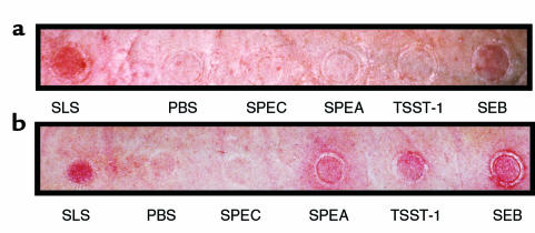
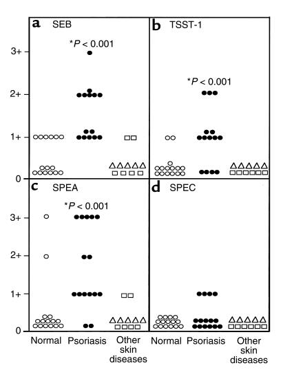
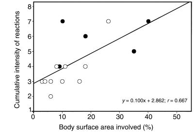
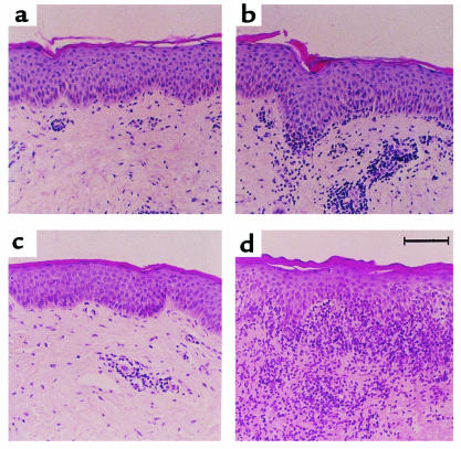
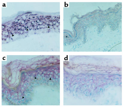
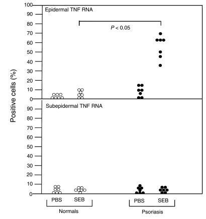
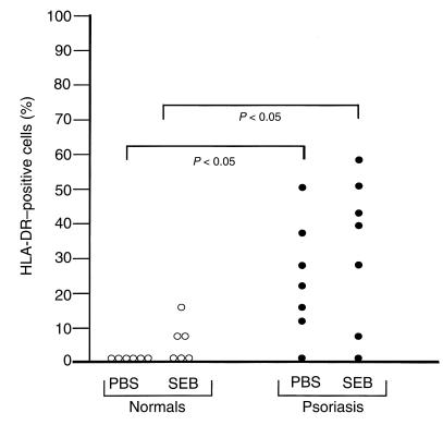
Streptococcal and staphylococcal superantigens (SAg's) have been implicated in the pathogenesis of inflammatory skin diseases, but the mechanisms by which these toxins act are unknown. The present study assessed the ability of nanogram quantities of topically applied purified toxic shock syndrome toxin-1 (TSST-1), staphylococcal enterotoxin type B, and streptococcal pyrogenic enterotoxin types A and C to induce inflammatory reactions in clinically uninvolved skin of normal controls and subjects with psoriasis, atopic dermatitis, and lichen planus. These SAg's triggered a significantly greater inflammatory skin response in psoriatics than in normal control subjects or in subjects with atopic dermatitis or lichen planus. Surprisingly, skin biopsies did not exhibit the T-cell receptor Vbeta stimulatory properties predicted for SAg-induced skin reactions. By 6 hours after patch testing with SAg's, TNF-alpha mRNA had increased in the epidermis (but not the dermis) in biopsies from psoriatics, compared with controls. Immunohistochemical studies revealed significantly higher HLA-DR expression in keratinocytes from psoriatics than from controls. However, a mutant TSST-1 protein that fails to bind HLA-DR did not elicit an inflammatory skin reaction. These results indicate that keratinocyte expression of HLA-DR enhances inflammatory skin responses to SAg's. They may also account for previous studies failing to demonstrate selective expansion of T-cell receptor Vbetas in psoriatics colonized with SAg-producing Staphylococcus aureus, and they identify a novel T cell-independent mechanism by which SAg's contribute to the pathogenesis of inflammatory skin diseases.


Figures

Figure 1
Figure 1
Typical examples of 48 hours of closed patch testing of exotoxins, PBS vehicle, and SLS on a normal subject (a) and a patient with psoriasis (b). Subjects underwent tape-stripping followed by closed patch testing of exotoxins SEB, TSST-1, SPEA, SPEC, PBS vehicle, or irritant SLS for 48 hours, as outlined in Methods. After removal of patches, the reactions were clinically rated as follows: 0 = no reaction; 1+ = macular erythema; 2+ = palpable erythema; 3+ = palpable erythema extending outside of the patch test site. These reactions were graded as follows: (a) SLS 1+, PBS 0, SPEC 0, SPEA 0, TSST-1 0, SEB 1+; (b) SLS 1+, PBS 0, SPEC 0, SPEA 3+, TSST-1 1+, SEB 2+.
Figure 2
Figure 2
Cutaneous reactivity of exotoxins on tape-stripped skin. Subjects with psoriasis, atopic dermatitis, lichen planus, or normal controls underwent our protocol of epidermal modification followed by closed patch testing. After removal of patches, the reactions were clinically rated as outlined in Figure 1. *P < 0.001 vs. normal controls.
Figure 3
Figure 3
Comparison of the cutaneous reactivity of psoriatics to exotoxins with extent of skin disease. At the time of closed patch testing, the amount of disease activity of psoriatic subjects was measured as percentage of body surface area involvement for the comparison. The cumulative clinical reactions to the 4 exotoxins tested were then compared with the percentage of body surface area involvement of psoriasis. The slope of the resulting curve was found to be statistically different from 0 (P < 0.05). Subjects who were found to develop psoriasis lesions in the sites of their skin biopsies (positive isomorphic phenomenon) are depicted by filled circles.
Figure 4
Figure 4
Histological evaluation of exotoxin-induced cutaneous reactions. Biopsies of reactions from subjects undergoing tape-stripping and closed patch testing for 48 hours were formalin-fixed, processed in paraffin, and stained with hematoxylin and eosin. (a) PBS reaction in a normal subject; (b) 1+ reaction to SEB in a normal subject; (c) PBS reaction in a psoriatic patient; and (d) 2+ reaction to SEB in a psoriatic patient.
Figure 5
Figure 5
Representative examples of TNF-α mRNA in situ hybridization (a and b) and HLA-DR immunostaining using APAAP technique (c and d). Shown in a is a representative example of TNF-α mRNA in the epidermis of a skin biopsy from a patient with psoriasis after 6 hours of SEB (×500); b is a skin section probed for TNF-α mRNA from a normal subject after 6 hours SEB (×500); c is an example of HLA-DR immunoreactivity in the epidermis of a patient with psoriasis after SEB application for 6 hours (×500); and d assesses HLA-DR immunoreactivity in the epidermis of a normal subject after SEB application for 6 hours.
Figure 6
Figure 6
Expression of TNF-α mRNA in PBS- and SEB-stimulated skin from patients with psoriasis and from normal controls. As described in Methods, subjects underwent tape-stripping followed by application of either SEB or PBS vehicle for 6 hours, and tissue samples were subjected to in situ hybridization to measure TNF-α gene expression by in situ hybridization.
Figure 7
Figure 7
Expression of HLA-DR in PBS- and SEB-stimulated skin from patients with psoriasis and from normal controls. As described in Methods, subjects underwent tape-stripping followed by application of either SEB or PBS vehicle for 6 hours, and tissue samples were subjected to immunohistochemistry to assess HLA-DR protein expression.

Comment in

References

    1. Nickoloff BJ. The cytokine network in psoriasis. Arch Dermatol. 1991;127:871–884. - PubMed
    1. Leung DYM, et al. Presence of IgE antibodies to staphylococcal exotoxins on the skin of patients with atopic dermatitis. Evidence for a new group of allergens. J Clin Invest. 1993;92:1374–1380. - PMC - PubMed
    1. Henderson CA, Highet AS. Acute psoriasis associated with Lancefield Group C and Group G cutaneous streptococcal infections. Br J Dermatol. 1988;118:559–561. - PubMed
    1. Leung DYM, Walsh P, Giorno R, Norris DA. A potential role for superantigens in the pathogenesis of psoriasis. J Invest Dermatol. 1993;100:225–228. - PubMed
    1. Leung DYM, et al. Evidence for a streptococcal superantigen-driven process in acute guttate psoriasis. J Clin Invest. 1995;96:2106–2112. - PMC - PubMed

Publication types

MeSH terms