. 1999 Nov;104(9):1199-211.
doi: 10.1172/JCI7615.

The chemokine SDF-1 stimulates integrin-mediated arrest of CD34(+) cells on vascular endothelium under shear flow

Affiliations

The chemokine SDF-1 stimulates integrin-mediated arrest of CD34(+) cells on vascular endothelium under shear flow

A Peled et al. J Clin Invest. 1999 Nov.

Abstract

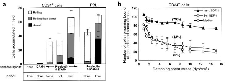
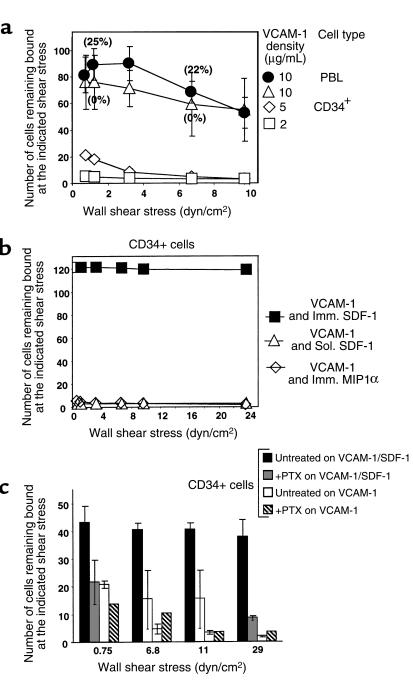
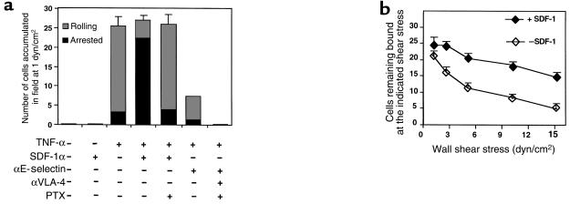
The chemokine SDF-1 plays a central role in the repopulation of the bone marrow (BM) by circulating CD34(+) progenitors, but the mechanisms of its action remain obscure. To extravasate to target tissue, a blood-borne cell must arrest firmly on vascular endothelium. Murine hematopoietic progenitors were recently shown in vivo to roll along BM microvessels that display selectins and integrins. We now show that SDF-1 is constitutively expressed by human BM endothelium. In vitro, human CD34(+) cells establish efficient rolling on P-selectin, E-selectin, and the CD44 ligand hyaluronic acid under physiological shear flow. ICAM-1 alone did not tether CD34(+) cells under flow, but, in the presence of surface-bound SDF-1, CD34(+) progenitors rolling on endothelial selectin rapidly developed firm adhesion to the endothelial surface, mediated by an interaction between ICAM-1 and its integrin ligand, which coimmobilized with SDF-1. Human CD34(+) cells accumulated efficiently on TNF-activated human umbilical cord endothelial cells in the absence of SDF-1, but they required immobilized SDF-1 to develop firm integrin-mediated adhesion and spreading. In the absence of selectins, SDF-1 also promoted VLA-4-mediated, Gi protein-dependent tethering and firm adhesion to VCAM-1 under shear flow. To our knowledge, this is the first demonstration that SDF-1 expressed on vascular endothelium is crucial for translating rolling adhesion of CD34(+) progenitors into firm adhesion by increasing the adhesiveness of the integrins VLA-4 and LFA-1 to their respective endothelial ligands, VCAM-1 and ICAM-1.


Figures

Figure 1
Figure 1
BM venules and capillaries reactive to anti–SDF-1 in human BM sections. SDF-1 immunostaining (arrows) of venous (a) and capillary (b) BM endothelium. ×100 (original magnification). No peroxidase staining of control mAb was observed on identical tissue sections.
Figure 2
Figure 2
Immunofluorescence flow cytometry of human CD34+ progenitors stained with antibodies to major vascular adhesion receptors and to the SDF-1 receptor CXCR4. (a) Cutaneous lymphocyte antigen (CLA) expression (detected by the HECA-452 mAb PSGL-1) and CD44 expression are shown. (b) Expression levels of the LFA-1 integrin CXCR4 and the integrin VLA-4. Negative control staining with nonbinding preimmune mouse IgG is shown in the filled histograms.
Figure 3
Figure 3
CD34+ cells roll on endothelial selectins and CD44 ligand under physiological shear flow. (a) Accumulation of CD34+ cells continuously perfused on substrates coated with E-selectin–IgG or P-selectin–IgG at stepwise incremented shear stresses; resistance of accumulated cells to detachment by elevated shear stresses. Cells were perfused for 45 seconds at 1 dyn/cm2, and then the flow was increased by stepwise increments every 5 seconds. The number of cells bound at the end of each interval of incremented shear stress was determined as described in Methods. Data points are presented as mean ± SD of 4 fields of view. Note that cells continued to attach to the substrate to a maximum shear stress of 3 dyn/cm2. Selectin-IgG fusion proteins were each coated at 4 μg/mL on substrate-immobilized protein A, yielding 130 sites/μm2 of fusion protein, corresponding to 260 selectin sites/μm2. (b) Velocities of CD34+ cells rolling at representative shear stresses on the P-selectin–IgG or E-selectin–IgG substrate as described in a. Each mean value represents a minimum of 15 cells ± SEM, determined in 2 fields of view. (c) Effect of shear stress on the CD44-mediated rolling of CD34+ cells tethered to immobilized hyaluronan. CD34+ cells were allowed to settle for 30 seconds on HA (coated at 1 mg/mL) and then subjected to a shear stress of 0.5 dyn/cm2 for 5 seconds, followed by 3 increments each of 0.5 dyn/cm2, 1 dyn/cm2, 2 dyn/cm2, and 3 dyn/cm2, with each increment lasting 5 seconds. The number of cells remaining adherent at the end of the indicated shear interval was determined in 4 representative fields and was expressed relative to the number of cells adhering to the HA-coated substrate at a shear stress of 2.5 dyn/cm2. The percentage of rolling cells within the adherent cells at low and high shear stresses is shown above the data points. The mean velocity of 20 CD34+ cells rolling on HA at representative shear stresses is shown in black squares below the graph. SEM of mean velocities determined at 6.5 dyn/cm2 and 9.5 dyn/cm2 were 2.8 μm/s and 2.7 μm/s, respectively. The data shown in ac are representative of 3 independent assays using CD34+ cells from different donors.
Figure 4
Figure 4
CD34+ cells and adult PBL express comparable levels of functional P-selectin ligand. (a) Accumulation of CD34+ cells and PBL perfused in identical numbers over immobilized P-selectin–IgG coated at 2 μg/mL. In control experiments, cells were briefly pretreated with the PSGL-1–blocking mAb KPL-1(αPSGL-1), or were perfused in a cation-free binding medium in the presence of EGTA. (b) Velocities of CD34+ cells and PBL at representative shear stresses on P-selectin–IgG or native (platelet-purified) P-selectin coated on substrates at densities supporting a comparable strength of adhesion of each cell type. Each mean value represents a minimum of 15 cells ± SEM, determined in 2 fields of view. *P < 0.002. **P < 0.016.
Figure 5
Figure 5
Tethering and rolling on P-selectin are prerequisites for SDF-1–triggered firm arrest of CD34+ cells on ICAM-1–containing surfaces. (a) CD34+ cell and T lymphocyte tethering to various adhesive substrates containing P-selectin, SDF-1, and ICAM-1 at a shear stress of 1 dyn/cm2. Motions of individual cells tethered to the different substrates were monitored over a 45-second period, and were divided into 3 categories as described in Methods. The fraction of each category within each experimental group (i.e., rolling, rolling-associated arrests, and immediate arrests) is presented in the stacked bars. The categories were analyzed only for cells that remained bound to the substrate at a shear stress of 2.5 dyn/cm2. (b) Resistance to detachment by incremented shear stresses of CD34+ cells accumulated at 1 dyn/cm2 on P-selectin/ICAM-1. The absolute numbers of cells accumulated during 1 minute at 1 dyn/cm2 and the number of cells remaining bound at the end of a 5-second interval of each shear increment are depicted. The percentage of stationary (arrested) cells within the cells remaining bound at a median shear stress (7.5 dyn/cm2) is shown in parentheses near the data points for each experimental group. ICAM-1 and SDF-1 were coated at 0.4 μg/mL and 10 μg/mL, respectively. P-selectin/ICAM-1 spots were prepared by mixing P-selectin and ICAM-1 in PBS/1% octyl glucoside and diluting the mixture in coating medium to final concentrations of 1 μg/mL and 0.5 μg/mL, respectively. Substrates were washed and then coated with SDF-1 as described in Methods. To assess the effect of soluble SDF-1, cells were preincubated in binding medium containing 1 μg/mL SDF-1 for 1 minute and perfused unwashed over the P-selectin/ICAM-1 substrate. Results shown in a and b are presented as mean of 2 determinations ± range. Sol., soluble; Imm., immobilized.
Figure 6
Figure 6
Immobilized SDF-1 stimulates CD34+ cell adhesion to VCAM-1. (a) Accumulation of CD34+ cells or PBL on sVCAM-1–coated substrates at 0.75 dyn/cm2, and resistance to detachment of accumulated cells by incremented shear stresses. (b) Effect of soluble (1 μg/mL) or immobilized SDF-1 or MIP-1α (co-coated at 2 μg/mL with sVCAM-1) on the accumulation of CD34+ cells at 0.75 dyn/cm2 and on the resistance of accumulated cells to detachment by elevated shear forces. (c) Inhibition by pertussis toxin (PTX) of SDF-1–triggered firm adhesion of CD34+ cells to sVCAM-1 alone or coated together with 2 μg/mL of SDF-1. In b and c, sVCAM-1 was coated at 2 μg/mL and 5 μg/mL, respectively.
Figure 7
Figure 7
CD34+ cell accumulation and development of firm adhesion on TNF-activated HUVEC under physiological shear flow. (a) Accumulation of CD34+ cells on intact and TNF-activated HUVEC; effect of EC-associated SDF-1 and blocking mAb’s against E-selectin or VLA-4. Cells were perfused at 1 dyn/cm2 for 45 seconds and then subjected to incremented shear stresses as described in Methods. The number of cells accumulated on the different HUVEC monolayers under the indicated experimental conditions was determined in 4 representative fields of view. Accumulated cells were divided into 2 categories: firmly arrested cells were cells that came to full arrest during accumulation at 1 dyn/cm2 and remained bound and stationary throughout the detachment assay (i.e., at a shear stress of 15 dyn/cm2). Rolling cells were defined as cells that continued to roll on the HUVEC monolayer immediately after tethering at 1 dyn/cm2, or cells that began to roll on the EC at elevated shear stresses and either remained adherent or moved from the field of view. The fractions of arrested or rolling cells among the cells initially accumulated in the field of view are shown in the stacked bars. (b) Resistance to detachment by shear flow of CD34+ cells accumulated at 1 dyn/cm2 on TNF-activated HUVEC. The absolute number of cells accumulated during 1 minute at 1 dyn/cm2 and the number of cells remaining bound at the end of each shear increment (each lasting 5 seconds) are depicted. Values are given as mean ± range of determinations in 4 fields of view. One of 4 independent experiments.

References

    1. Vermeulen M, et al. Role of adhesion molecules in the homing and mobilization of murine hematopoietic stem and progenitor cells. Blood. 1998;92:894–900. - PubMed
    1. Quesenberry PJ, Becker PS. Stem cell homing: rolling, crawling, and nesting. Proc Natl Acad Sci USA. 1998;95:15155–15157. - PMC - PubMed
    1. Verfaillie CM. Adhesion receptors as regulators of the hematopoietic process. Blood. 1998;92:2609–2612. - PubMed
    1. Mazo IB, et al. Hematopoietic progenitor cell rolling in bone marrow microvessels: parallel contributions by endothelial selectins and vascular cell adhesion molecule 1. J Exp Med. 1998;188:465–474. - PMC - PubMed
    1. Frenette PS, Subbarao S, Mazo IB, von Andrian UH, Wagner DD. Endothelial selectins and vascular cell adhesion molecule-1 promote hematopoietic progenitor homing to bone marrow. Proc Natl Acad Sci USA. 1998;95:14423–14428. - PMC - PubMed

Publication types