. 1999 Nov;104(9):1235-44.
doi: 10.1172/JCI7377.

Dilated cardiomyopathy in homozygous myosin-binding protein-C mutant mice

Affiliations

Dilated cardiomyopathy in homozygous myosin-binding protein-C mutant mice

B K McConnell et al. J Clin Invest. 1999 Nov.

Erratum in

  • J Clin Invest 1999 Dec;104(12):1771. Fischman DH [corrected to Fischman DA]

Abstract

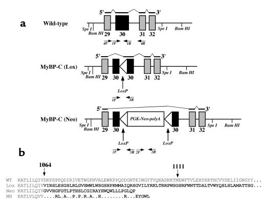
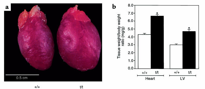
To elucidate the role of cardiac myosin-binding protein-C (MyBP-C) in myocardial structure and function, we have produced mice expressing altered forms of this sarcomere protein. The engineered mutations encode truncated forms of MyBP-C in which the cardiac myosin heavy chain-binding and titin-binding domain has been replaced with novel amino acid residues. Analogous heterozygous defects in humans cause hypertrophic cardiomyopathy. Mice that are homozygous for the mutated MyBP-C alleles express less than 10% of truncated protein in M-bands of otherwise normal sarcomeres. Homozygous mice bearing mutated MyBP-C alleles are viable but exhibit neonatal onset of a progressive dilated cardiomyopathy with prominent histopathology of myocyte hypertrophy, myofibrillar disarray, fibrosis, and dystrophic calcification. Echocardiography of homozygous mutant mice showed left ventricular dilation and reduced contractile function at birth; myocardial hypertrophy increased as the animals matured. Left-ventricular pressure-volume analyses in adult homozygous mutant mice demonstrated depressed systolic contractility with diastolic dysfunction. These data revise our understanding of the role that MyBP-C plays in myofibrillogenesis during cardiac development and indicate the importance of this protein for long-term sarcomere function and normal cardiac morphology. We also propose that mice bearing homozygous familial hypertrophic cardiomyopathy-causing mutations may provide useful tools for predicting the severity of disease that these mutations will cause in humans.


Figures

Figure 1
Figure 1
Schematic of (a) the genomic structure of wild-type MyBP-C, MyBP-C(LoxP), and MyBP-C(Neo) alleles, the RNAs produced by each allele, and (b) the structure of the MyBP-C proteins encoded by each allele. (a) Exons 29–32 of the murine cardiac MyBP-C gene are shown for each allele (see Methods). The mutations alter only exon 30; all other exons are identical. The black lines above each gene segment reflect the structure of the encoded RNA indicated in the 5′→3′ orientation; thick lines are exons incorporated into the RNA, thin lines indicate skipped segments of the gene not found in RNA. The genotypes of mice were determined with primers 1F, 1R, 2F, 2R, and 3R. Primers 4F and 4R were used to determine the structure of MyBP-C RNA; structures of the deduced RNAs are indicated above each allele. (b) The structure of carboxyl ends of the MyBP-C polypeptides encoded by MyBP-C(Neo) and MyBP-C(Lox) alleles were deduced from the sequences of RNA found in the left ventricles of homozygous mice bearing the indicated allele. The residues (between residues 1064 and 1111) of the wild-type protein are encoded by exon 30. Novel amino acid residues at the carboxyl end of the mutant proteins (bold) are encoded by altered reading of exon 30 (Lox) or exon 31 (Neo). The wild-type MyBP-C protein is 1270 amino acids whereas the MyBP-C(Lox) protein is 1240-amino acids long; the complete sequences of these 2 carboxyl ends are not shown. The carboxyl end of the analogous human mutation found in family NN (3) is shown for comparison. Differences between the family NN protein and the MyBP-C(Neo) protein reflect sequence differences between the mouse and human MyBP-C gene exon 31.
Figure 2
Figure 2
MyBP-C mRNA and protein expression levels in hearts of 12-week-old homozygous MyBP-C(Neo) mice (designated t/t). (a) Northern blot analyses of RNAs from left ventricle of wild-type (+/+) and MyBP-C(Neo) mice hybridized with murine MyBP-C and GAPDH probes. (b) Western blot analyses identified the 150-kDa MyBP-C protein in myofibrillar extracts of the LV of wild-type (+/+) and homozygous MyBP-C(Neo) mice (t/t).
Figure 3
Figure 3
Comparison of hearts from 8–12-week-old wild-type and homozygous mutant mice. (a) Gross morphology of homozygous MyBP-C(Neo) (designated t/t) and wild-type (+/+) hearts and (b) heart-to-body weight or LV-to-body weight ratios demonstrate significant increases (P < 0.001) in cardiac size of homozygous mutant mice (t/t) versus wild-type mice (+/+).
Figure 4
Figure 4
Histology of 8–12-week-old wild-type (a) and MyBP-C(Neo) (bd) mouse hearts. Sections were stained with H&E (a and c) or Masson trichrome (b and d).
Figure 5
Figure 5
Transmission electron micrographs of sarcomeres from 12-week-old wild-type (a) and MyBP-C(Neo) mouse hearts (t/t). Note the unusual appearance of the M-line (asterisk) in MyBP-C(Neo) mouse-derived sarcomeres. Bar, 0.5 μm.
Figure 6
Figure 6
Cardiac function in wild-type and MyBP-C(Neo) mice. (ac) Schematic and M-mode echocardiographic tracings from 12-week-old wild-type (b) and MyBP-C(Neo) mice (c). LV dimensions are calculated from measurements of anterior wall (1), posterior wall (2), end-diastolic diameter (3), and end-systolic diameter (4). Increased contrast in the posterior papillary muscle probably reflects calcification (c) (arrow). (d) Pressure volume relations and dP/dt profiles of wild-type (+/+) and MyBP-C(Neo) mice (t/t).
Figure 7
Figure 7
Altered gene expression in hearts from MyBP-C(Neo) mice. Northern blots (a) show increased expression of α-skeletal actin and BNP mRNAs in comparison with GAPDH transcripts. Silver-stained SDS-page gels (b) demonstrate altered expression of cardiac MHC isoforms in total ventricular extracts from MyBP-C(Neo) mouse hearts (from 8–12-week-old animals; designated t/t) compared with protein from wild-type mice (+/+).

References

    1. Chien, K.R., Grace, A.A., and Hunter, J.J. 1999. Molecular and cellular biology of cardiac hypertrophy and failure. In Molecular basis of cardiovascular disease. K.R. Chien, editor. W.B. Saunders Co. Philadelphia, PA. 251–264.
    1. Rodkey, S.M., Ratliff, N.B., and Young, J.B. 1998. Cardiomyopathy and myocardial failure. In Comprehensive cardiovascular medicine. E.J. Topal, editor. Lippincott-Raven Publishers. Philadelphia, PA. 2589–2620.
    1. Watkins H, et al. Mutations in the cardiac myosin binding protein C gene on chromosome 11 cause familial hypertrophic cardiomyopathy. Nat Genet. 1995;11:434–437. - PubMed
    1. Bonne G, et al. Cardiac myosin binding protein-C gene splice acceptor site mutation is associated with familial hypertrophic cardiomyopathy. Nat Genet. 1995;11:438–440. - PubMed
    1. Niimura H, et al. Mutations in the gene for cardiac myosin-binding protein C and late-onset familial hypertrophic cardiomyopathy. N Engl J Med. 1998;338:1248–1257. - PubMed

Publication types

MeSH terms