. 1999 Nov;104(9):1307-17.
doi: 10.1172/JCI6688.

Immune-mediated inflammation directly impairs pulmonary function, contributing to the pathogenesis of Pneumocystis carinii pneumonia

Affiliations

Immune-mediated inflammation directly impairs pulmonary function, contributing to the pathogenesis of Pneumocystis carinii pneumonia

T W Wright et al. J Clin Invest. 1999 Nov.

Abstract

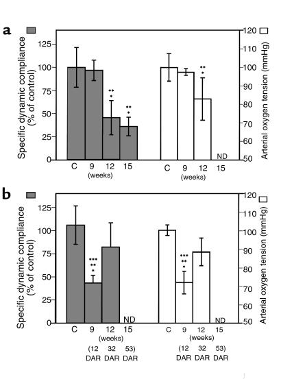
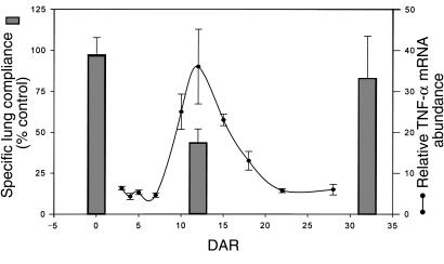
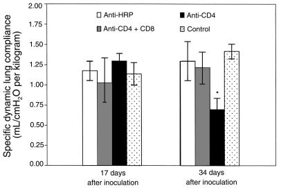
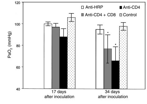
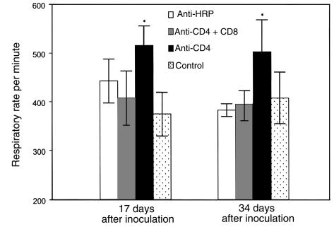
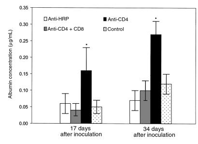
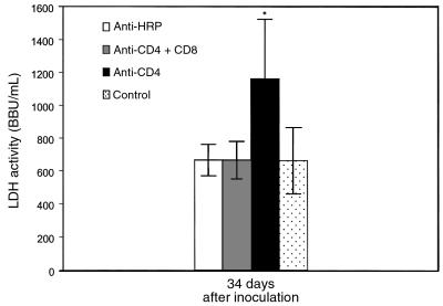
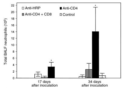
The clinical severity of Pneumocystis carinii pneumonia (PCP) correlates closely with the appearance of pulmonary markers of inflammation. Therefore, a model system was developed whereby physiological studies could be performed on live mice to determine the extent to which pulmonary inflammation contributes to respiratory impairment during PCP. P. carinii-infected severe combined immunodeficient mice displayed little evidence of pulmonary inflammation and exhibited normal oxygenation and dynamic lung compliance. When comparably infected littermates were immunologically reconstituted, however, an intense immune-mediated inflammatory response was observed that resulted in significant decreases in both lung compliance and oxygenation. As the pneumonia resolved pulmonary function returned toward normal. To begin to define the cell populations contributing to inflammation-associated respiratory impairment during PCP, similar studies were performed in CD4(+) T cell-depleted mice. Mice depleted of both CD4(+) and CD8(+) cells developed infection, but they demonstrated neither abnormal lung compliance nor increased respiratory rate and displayed no markers of lung injury. In contrast, mice depleted of only CD4(+) T cells exhibited severe pulmonary inflammation and injury, decreased oxygenation and lung compliance, and increased respirations. Respiratory compromise was associated with the presence of activated CD8(+) cells and neutrophils in broncho-alveolar lavage fluid. These observations provide direct experimental evidence that the host's response to P. carinii directly impairs pulmonary function and contributes to the pathogenesis of PCP. Furthermore, CD8(+) T cells likely contribute to the respiratory compromise observed during PCP.


Figures

Figure 1
Figure 1
Impaired pulmonary function during PCP in SCID mice. For comparison of nonreconstituted and reconstituted SCID mice, time points are expressed in weeks after exposure to P. carinii. (a) Progressive decline in specific lung compliance and PaO2 in nonreconstituted P. carinii–infected SCID mice. (b) Respiratory impairment and recovery after immune reconstitution of P. carinii–infected SCID mice. ND, not determined. C, P. carinii–free control. *P < 0.05 vs. P. carinii–free controls. **P < 0.05 vs. P. carinii–infected SCID mice at 9 weeks after exposure. ***P < 0.05 vs. reconstituted P. carinii–infected SCID mice at 32 DAR.
Figure 2
Figure 2
Temporal relationship of pulmonary inflammation and specific dynamic lung compliance after immune reconstitution of P. carinii–infected SCID mice. Dynamic lung compliance and pulmonary TNF-α mRNA abundance were measured at various times after reconstitution. The time course of TNF-α mRNA abundance has been reported previously (10) and serves as a reliable marker of pulmonary inflammation in this model. Pulmonary inflammation is minimal and lung compliance is normal in infected mice at 0 DAR. However, during the peak inflammatory phase after reconstitution (12 DAR), lung compliance is dramatically decreased. Finally, by 32 DAR the infection has been cleared, pulmonary inflammation is resolving, and lung compliance measurements approach normal.
Figure 3
Figure 3
Effect of CD8+ T cells on dynamic lung compliance during PCP in mice. Specific dynamic lung compliance measurements were taken on CD4-depleted, CD4/CD8–depleted, and anti-HRP–treated mice inoculated with P. carinii 17 or 34 days previously. Controls were uninfected C57BL/6 mice. Values are mean ± 1 SD (n = 5). *P < 0.05 vs. CD4/CD8–depleted, anti-HRP–treated, and uninfected mice at 17 and 34 days after inoculation.
Figure 4
Figure 4
Effect of CD8+ T cells on arterial oxygenation during PCP in mice. PaO2 was measured in CD4-depleted, CD4/CD8–depleted, and anti-HRP–treated mice inoculated with P. carinii 17 or 34 days previously. Controls were uninfected C57BL/6 mice. Values are mean ± 1 SD (n = 5). *P < 0.05 vs. anti-HRP–treated and uninfected mice at 34 days after inoculation.
Figure 5
Figure 5
Effect of CD8+ T cells on respiratory rates during PCP in mice. Respiratory rates were measured in CD4-depleted, CD4/CD8–depleted, and anti-HRP–treated mice inoculated with P. carinii 17 or 34 days previously. Controls were uninfected C57BL/6 mice. Values are mean ± 1 SD (n = 5). *P < 0.05 vs. CD4/CD8–depleted, anti-HRP–treated, and uninfected mice at days 17 and 34 after inoculation.
Figure 6
Figure 6
Effect of CD8+ T cells on BALF albumin concentration during PCP in mice. Albumin content was measured in the BALF of CD4-depleted, CD4/CD8–depleted, and anti-HRP–treated mice inoculated with P. carinii 17 or 34 days previously. Controls were uninfected C57BL/6 mice. Values are mean ± 1 SD (n = 5). *P < 0.05 vs. CD4/-CD8–depleted, anti-HRP–treated, and uninfected mice at 17 and 34 days after inoculation.
Figure 7
Figure 7
Effect of CD8+ T cells on serum LDH activity during PCP in mice. LDH activity in the serum of CD4-depleted, CD4/CD8–depleted, and anti-HRP–treated mice infected with P. carinii 34 days previously was measured. Controls were uninfected C57BL/6 mice. Values are mean ± 1 SD (n = 5). *P < 0.05 vs. CD4/CD8–depleted, anti-HRP–treated, and uninfected mice at 34 days after inoculation.
Figure 8
Figure 8
Effect of CD8+ T cells on pulmonary neutrophil recruitment during PCP in mice. Neutrophils were quantified in the BALF of CD4-depleted, CD4/CD8–depleted, and anti-HRP–treated mice infected with P. carinii 17 or 34 days previously. Controls were uninfected C57BL/6 mice. Values are mean ± 1 SD (n = 5). *P < 0.05 vs. CD4/CD8-depleted, anti-HRP–treated, and uninfected mice at 17 and 34 days after inoculation.

References

    1. Su TH, Martin WJ., II Pathogenesis of host response in Pneumocystis carinii pneumonia. Annu Rev Med. 1994;45:261–272. - PubMed
    1. Sepkowitz KA. Pneumocystis carinii pneumonia in patients without AIDS. Clin Infect Dis. 1993;17(Suppl. 2):S41–S422. - PubMed
    1. CDC. Guidelines for prophylaxis against Pneumocystis carinii pneumonia for persons infected with Human Immunodeficiency Virus. MMWR Morb Mortal Wkly Rep. 1989;38(Suppl. 5):1–9. - PubMed
    1. Kovacs JA, et al. Pneumocystis carinii pneumonia: a comparison between patients with the acquired immunodeficiency syndrome and patients with other immunodeficiencies. Ann Intern Med. 1984;100:663–671. - PubMed
    1. Benfield TL, et al. Prognostic value of interleukin-8 in AIDS-associated Pneumocystis carinii pneumonia. Am J Respir Crit Care Med. 1995;151:1058–1062. - PubMed

Publication types