. 1999 Nov;155(5):1525-33.
doi: 10.1016/S0002-9440(10)65468-5.

Hepatocarcinoma-intestine-pancreas/pancreatic associated protein (HIP/PAP) is expressed and secreted by proliferating ductules as well as by hepatocarcinoma and cholangiocarcinoma cells

Affiliations

Hepatocarcinoma-intestine-pancreas/pancreatic associated protein (HIP/PAP) is expressed and secreted by proliferating ductules as well as by hepatocarcinoma and cholangiocarcinoma cells

L Christa et al. Am J Pathol. 1999 Nov.

Abstract

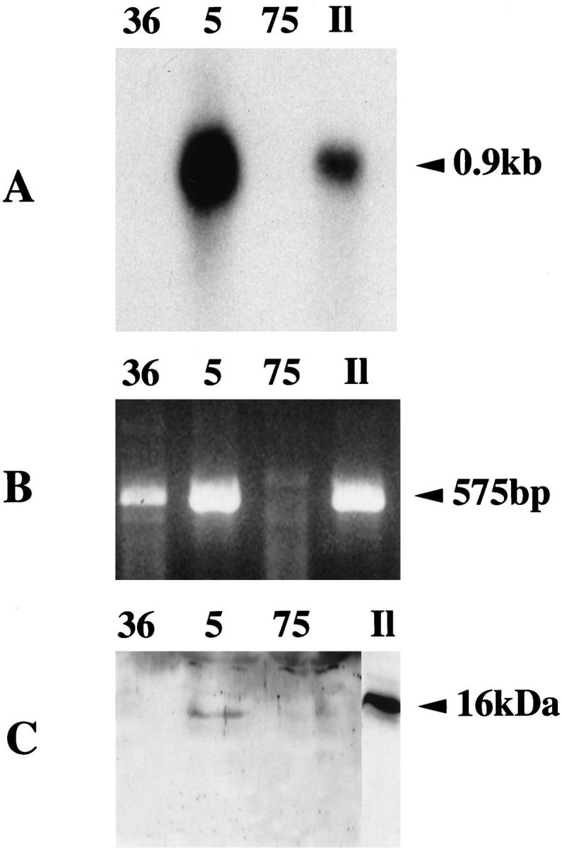
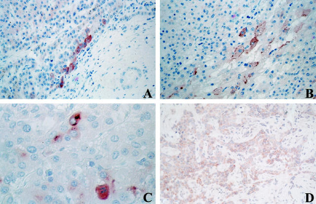
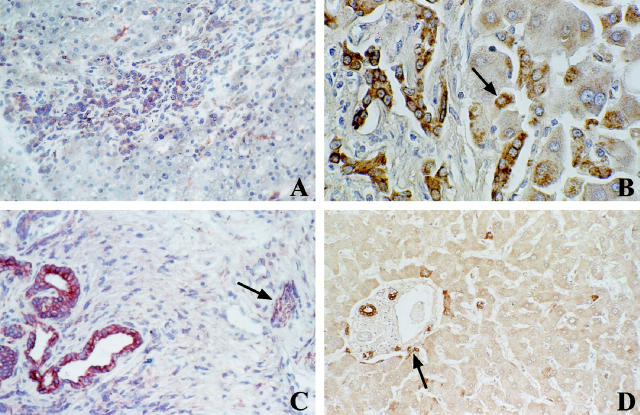
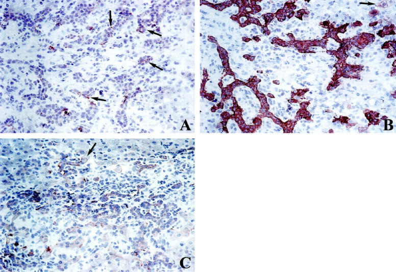
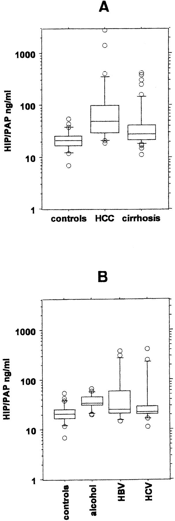
Hepatocarcinoma-intestine-pancreas/pancreatic associated protein (HIP/PAP) gene was identified because of its increased expression in 25% of human hepatocellular carcinoma. HIP/PAP protein, a C-type lectin, binds laminin, acts as an adhesion molecule for hepatocytes, and has also been described as an acute phase secretory protein during acute pancreatitis in humans and rats. We investigated HIP/PAP protein expression in patients with various liver diseases associated with ductular reaction. At the same time, we analyzed patients with hepatocellular carcinoma and cholangiocarcinoma, and tested HIP/PAP protein levels in sera to establish the pattern of secretion. Our data show that HIP/PAP expression was not restricted to hepatocellular carcinoma, but was also detected in cholangiocarcinoma cells as well as in reactive non-malignant bile ductules. In contrast, HIP/PAP protein expression was undetectable in normal mature hepatocytes, but some ductular cells localized at the interface of portal tracts with parenchyma were HIP/PAP immunoreactive in normal liver. Finally, we present evidence that HIP/PAP serum levels were increased in 21/28 (75%) patients with hepatocellular carcinoma, and in 25/51 (49%) patients with nonmalignant cirrhosis. Altogether, these results suggest that HIP/PAP protein may be implicated in hepatocytic and cholangiolar differentiation and proliferation.


Figures

Figure 1.
Figure 1.
Northern blot 20 μg (A) and RT-PCR 1 μg (B) analysis of tumoral liver RNA from patients 36, 5, 75, and normal human ileum RNA (Il). Patient 36 was negative by Northern blot and positive by RT-PCR. Patient 5 was positive by Northern blot and RT-PCR. Patient 75 was negative by Northern blot and RT-PCR. Western blot analysis of serum (10 μl) from patients 36, 5, 75, and control normal human ileum tissue (20 μg) (Il) (C). HIP/PAP serum levels were 19, 237, and 42 ng/ml in patients 36, 5, and 75, respectively, as quantified using an ELISA test. Only patient 5 gave a detectable signal.
Figure 2.
Figure 2.
Immunolocalization of HIP/PAP protein in HCC (A-C) and cholangiocarcinoma (D). A: Immunolocalization of HIP/PAP protein at the frontier between affected (left) and non-affected portions of the liver section (HCC no. 81, paraffin section, pre-HIP antibodies, original magnification, ×250). B: Intracytoplasmic brown granules occupying most of the cytoplasm, plus plasmic membrane labeling (HCC no. 82, paraffin section, pre-HIP antibodies, original magnification, ×250). C: Cytoplasmic granulous labeling plus perinuclear bodies (patient 93, paraffin section, pre-HIP antibodies, original magnification, ×400). D: Cholangiocarcinoma with a majority of tumor cells showing varying degrees of cytoplasmic immunoreactivity for HIP/PAP (frozen section, pre-HIP antibodies, original magnification, ×250).
Figure 3.
Figure 3.
Immunolocalization of HIP/PAP protein in nonmalignant tissues. A: Chronic extrahepatic subobstruction. Reactive bile ductules at the edge of a portal tract are immunoreactive for HIP/PAP in their cytoplasm. (frozen section, pre-HIP antibodies, original magnification, ×250). B: Active cirrhosis. Reactive ductules and hepatocyte-like cells (arrow) are immunoreactive for HIP/PAP (paraffin section, CRD antibodies, original magnification, ×400). C: Chronic active hepatitis C. Larger portal tract showing immunoreactivity for HIP/PAP in ductular structures (left) and in nerve bundles (arrow) (frozen section, pre-HIP antibodies, original magnification, ×250). D: Portal tract in normal liver. Immunoreactivity in bile ducts and in some scarce ductular cells (arrow) with negative surrounding hepatocytes (paraffin section, CRD antibodies, original magnification, ×250).
Figure 4.
Figure 4.
Semiserially cut cryostat sections of a portal tract in an extrahepatic subobstruction, immunostained (A) for HIP/PAP in a subpopulation of ductular cells with varying intensity (arrows), original magnification, ×250, (B) for cytokeratin 7 (bile duct type cytokeratin) in reactive ductules with a strong positive labeling. Some periportal hepatocyte-like intermediate cells are also immunoreactive (arrow), original magnification, ×250, (C) for OV6. The same portal tract shows immunoreactivity for OV-6 in reactive ductules and in hepatocyte-like intermediate cells (arrow), original magnification, ×250.
Figure 5.
Figure 5.
Serum HIP/PAP levels. Each box plot comprises five horizontal lines displaying the 10th, 25th, 50th, 75th, and 90th percentiles of a variable. All values for the variable above the 90th percentile and below the 10th percentile are plotted separately, so that the box plots are valuable in highlighting any outliers. A: Serum HIP/PAP concentrations were determined in controls (n = 26), in patients with HCC (n = 28), and in patients with cirrhosis without overt HCC (n = 51). B: Serum HIP/PAP concentrations were determined in controls (n = 26) and in cirrhotic patients taking into account the etiology of the cirrhosis (alcohol n = 17, HBV = 17, and HCV = 16). One cirrhotic patient with primitive sclerosing cholangitis (HIP/PAP = 111 ng/ml) was excluded.

References

    1. Lasserre C, Christa L, Simon MT, Vernier P, Bréchot C: A novel gene (HIP) activated in human liver cancer. Cancer Res 1992, 52:5089-5095 - PubMed
    1. Drickamer K: Calcium-dependent carbohydrate-recognition domains in animal proteins. Curr Opin Struct Biol 1993, 3:393-400
    1. Frigerio JM, Dusetti NJ, Garrido P, Dagorn JC, Iovanna JL: The pancreatitis associated protein III (PAPIII), a new member of the PAP gene family. Biophys Biophys Acta 1993, 1216:329-331 - PubMed
    1. Narushima Y, Unno M, Nakagawa KI, Mori M, Miyashita H, Suzuki Y, Nogushi N, Takasawa S, Kumagai T, Yonekura H, Okamoto H: Structure, chromosomal localization, and expression of mouse genes encoding type III Reg, Reg IIIa, Reg IIIb, Reg IIIc. Gene 1997, 185:159-168 - PubMed
    1. Itoh T, Teraoka H: Cloning and tissue-specific expression of cDNAs for the human and mouse homologues of rat pancreatitis-associated protein (PAP). Biophys Biophys Acta 1993, 1172:184-186 - PubMed

Publication types

MeSH terms