. 1999 Nov;155(5):1577-86.
doi: 10.1016/S0002-9440(10)65474-0.

Stromal-cell derived factor is expressed by dendritic cells and endothelium in human skin

Affiliations

Stromal-cell derived factor is expressed by dendritic cells and endothelium in human skin

J L Pablos et al. Am J Pathol. 1999 Nov.

Abstract

Stromal-cell derived factor or SDF-1 is a CXC chemokine constitutively expressed by stromal bone marrow cell cultures that binds to the G-protein-coupled receptor CXCR4. SDF-1/CXCR4 represents a unique, nonpromiscuous ligand/receptor pair that plays an essential role in prenatal myelo- and lymphopoiesis as well as in cardiovascular and neural development. SDF-1 prevents entry of CXCR4-dependent (X4) HIV viruses in T lymphocytes, by binding and internalizing CXCR4. The expression pattern of SDF-1 protein in normal tissues is not known. Here we describe an analysis of SDF-1 mRNA and protein in normal and inflamed skin by in situ hybridization and immunohistochemistry, using a novel anti-SDF-1 monoclonal antibody. We also describe the expression pattern of CXCR4 receptor by immunohistochemistry. Our results show that SDF-1 protein and mRNA are normally expressed by endothelial cells, pericytes, and either resident or explanted CD1a+ dendritic cells. Epithelial cells of sweat glands but not keratinocytes also express SDF-1. In various inflammatory skin diseases, a large number of mononuclear cells and fibroblasts in close contact with CXCR4-positive lymphocytic infiltrates also express SDF-1. CXCR4 was also detected in many different normal cell types, including endothelial and epithelial cells, which points to a role for SDF-1/CXCR4 cell signaling in vascular and epithelial homeostasis. The demonstration of SDF-1 expression in dendritic and endothelial cells provides new insights into the mechanisms of normal and pathological lymphocyte circulation and makes it possible to envisage a role for locally secreted SDF-1 in the selective incapacity of mucosal dendritic cells to support and propagate infection by X4 HIV isolates.


Figures

Figure 1.
Figure 1.
Expression and detection of SDF-1 gene products in mammalian cells by the K15C MAb. a: TIGEF cells were transfected with pCDNA3, SDF-1α, or SDF-1β. Forty-eight hours after transfection cell supernatants (pcDNA, SDF-1α, or SDF-1β) were collected, and their ability to promote CXCR4 down-regulation from the surface of lymphoblastoid Jurkat T cells was assessed. Supernatants from untransfected TIGEF cell cultures (Untr.) were used as controls. The expression of CXCR4 was analyzed by cytofluorometry, using the CXCR4-antibody 12G5 or an isotype-matched mouse immunoglobulin (Ctrl). b: TIGEF cells were transfected as indicated above and treated or not for the last 4 hours in culture with Brefeldine A (Bref. A). Cells were collected 48 hours after transfection, and cytoplasmic proteins (60 μg) were analyzed by Western blot with the K15C antibody. The ability of the K15C mAb to recognize TIGEF-expressed or synthetic (100, 50, or 25 ng) SDF-1 isoforms was compared.
Figure 2.
Figure 2.
Detection by confocal microscopy of the intracellular accumulation of SDF-1β by the K15C mAb. TIGEF cells cotransfected with SDF-1β (or an insertless pcDNA3 plasmid) and GFP (green) expression vectors were incubated with K15C mAb and a secondary antibody conjugated to cye-3 (red). Cultures were treated with Brefeldine A. A and B: Cotransfection of SDF-1β and GFP expression vectors. C and D: Cotransfection of insertless pcDNA3 and a GFP expression vector. A and C: Simultaneous detection of GFP (green) and SDF-1β (red). B and D: Detection of SDF-1β (red).
Figure 3.
Figure 3.
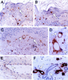
SDF-1 immunostaining in normal skin. A: Epidermal scattered DC were immunostained by anti-SDF-1 K15C MAb (arrow). B: A parallel section shows the identical morphology and distribution of SDF-1-positive and CD1a-positive epidermal DC (Langerhans cells). C: Dermis also contains scattered SDF-1 immunostained DC (arrow). EC and pericytes of dermal capillary vessels (arrowhead) were also immunostained by anti-SDF-1 MAb.D: Both EC (arrow) and pericytes (arrowhead) of small capillary vessels were immunostained. E: EC of larger vessels displayed weak immunostaining with anti-SDF-1 MAb. F: Epithelial cells of sweat glands showed strong SDF-1 immunostaining (arrow). Sweat gland immunostaining was not uniform, and there were unstained areas (arrowhead).
Figure 4.
Figure 4.
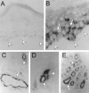
SDF-1 mRNA expression by ISH in normal skin. A: SDF-1 sense riboprobes did not produce any signal above background (arrowheads mark the dermoepidermal union). B: SDF-1 mRNA expression was detected in epidermal Langerhans cells (marked by arrows). C: Both small capillary vessels (arrow) and larger blood vessels (arrowhead) displayed EC labeling. D: A small capillary vessel with higher magnification shows labeling of both EC (arrow) and pericytes (arrowhead). E: Sweat gland epithelial cells showed uneven mRNA SDF-1 labeling, with alternating labeled (arrow) and unlabeled (arrowhead) areas.
Figure 5.
Figure 5.
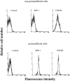
Expression of SDF-1 and CXCR4 in purified CD1a+ DC explanted from skin. Cytofluorometric analysis of CD1a+ cells labeled with the anti-SDF K15C mAb, 12G5 anti-CXCR4 mAb, or isotype-matched immunoglobulin (control). SDF-1 was only detected in permeabilized cells. CXCR4 was present at low levels on the surface of nonpermeabilized cells, and its detection increased in permeabilized cells.
Figure 6.
Figure 6.
Detection of SDF-1 mRNA in CD1a+ DC explanted from the skin. Foreskin fibroblasts and PBMC were used, respectively, as sources of positive and negative controls for the expression of SDF-1 gene. cDNAs were generated by reverse transcription (RT) of total RNA and amplified by PCR. Aliquots of each sample were similarly processed for the expression of β-actin, which was used as a cDNA loading control. To rule out DNA contamination and verify the specificity of the PCR-amplified signal, addition of reverse transcriptase was omitted where indicated (RT).
Figure 7.
Figure 7.
SDF-1 and CXCR4 immunostaining in inflammatory infiltrates from skin autoimmune diseases. A, C: SDF-1 immunostaining is present in a large number of fibroblasts (arrows) and large mononuclear cells (arrowheads) but not in small lymphocytes of inflammatory infiltrates. B, D: Virtually all lymphocytes and mononuclear cells within inflammatory infiltrates were CXCR4 positive. A, C: Cutaneous lupus. B, D: Dermatomyositis.
Figure 8.
Figure 8.
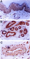
CXCR4 immunostaining in normal skin. A: Normal epidermal keratinocytes were uniformly stained by anti-CXCR4 antibodies. EC and pericytes (arrowhead) and dermal fibroblasts (arrow) were also immunostained. B: Normal sweat gland epithelial cells were strongly and uniformly immunostained. C: Larger vessels also showed strong CXCR4 staining (arrowhead). Cells with dendritic (arrow) and fibroblastic morphology around blood vessels are also immunostained.

References

    1. Baggiolini M, Dewald B, Moser B: Human chemokines: an update. Annu Rev Immunol 1997, 15:675-705 - PubMed
    1. Nagasawa T, Tachibana K, Kawabata K: A CXC chemokine SDF-1/PBSF. A ligand for a HIV receptor, CXCR4. Adv Immunol 1999, 71:211-228 - PubMed
    1. Shirozu M, Nakano T, Inazawa J, Tashiro K, Tada H, Shinohara T, Honjo T: Structure and chromosomal localization of the human stromal cell-derived factor 1 (SDF-1) gene. Genomics 1995, 28:495-500 - PubMed
    1. Nagasawa T, Kikutani H, Kishimoto T: Molecular cloning and structure of a pre-B-cell growth-stimulating factor. Proc Natl Acad Sci USA 1994, 91:2305-2309 - PMC - PubMed
    1. Nagasawa T, Hirota S, Tachibana K, Tachibana K, Takakura N, Nishikawa S, Kitamura Y, Yoshida N, Kikutani H, Kishimoto T: Defects of B-cell lymphopoiesis and bone-marrow myelopoiesis in mice lacking the CXC chemokine PBSF/SDF-1. Nature 1996, 382:635-638 - PubMed

Publication types

LinkOut - more resources