

doi: 10.1016/S0002-9440(10)65483-1.
Nephrin localizes at the podocyte filtration slit area and is characteristically spliced in the human kidney
Affiliations
- PMID: 10550324
- PMCID: PMC1866978
- DOI: 10.1016/S0002-9440(10)65483-1
Item in Clipboard
Nephrin localizes at the podocyte filtration slit area and is characteristically spliced in the human kidney
Am J Pathol.
1999 Nov.
Abstract
Defects in the newly reported gene NPHS1 in chromosome 19 cause the massive proteinuria of Finnish type congenital nephrotic syndrome (CNF). Together with its gene product, nephrin, NPHS1 is providing new understanding of the pathophysiological mechanisms of glomerular filtration. Here we show the characteristic splicing of NPHS1 mRNA in the normal and CNF kidneys and localize nephrin exclusively in the glomerulus and to the filtration slit area by light and immunoelectron microscopy. These results indicate that nephrin is a new protein of the interpodocyte filtration slit area with a profound role in the pathophysiology of the filtration barrier.
Figures
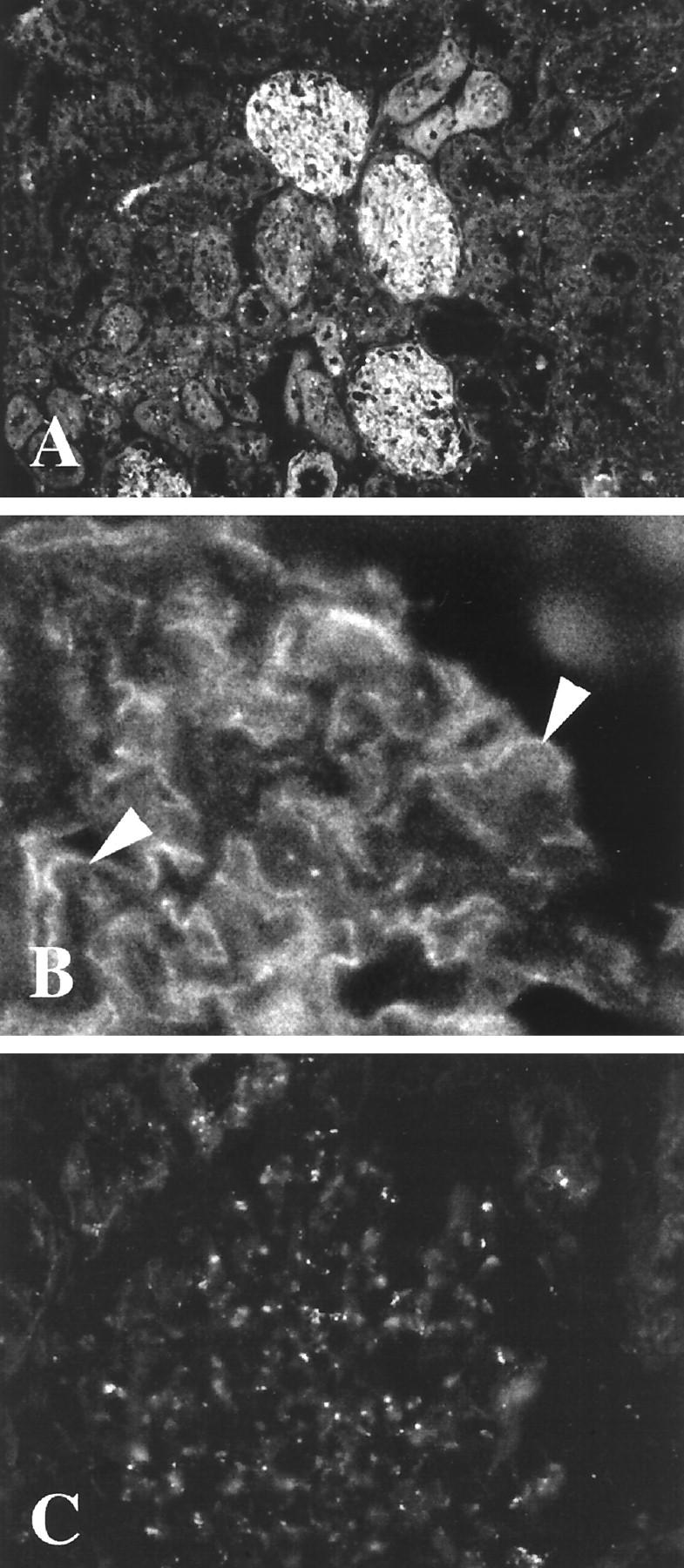
A, B: Frozen section of normal human kidney stained with antipeptide antibodies to the intracellular nephrin-specific sequence in immunofluorescence. Note the reactivity exclusively in glomerulus (A) as finely granular stretches of linear reactivity (arrows in B) in a pattern typical for podocytes. Reactivity of preimmune serum is given in C. Original magnifications, ×180 (A) and ×360 (B and C)
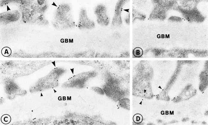
Localization of nephrin in the glomerular capillary wall of normal human kidney (A-D). Labeling is found in association with the slit diaphragms in filtration slits. In addition, small clusters of nephrin are also seen on the surface of podocytes (large arrowheads) as well as occasionally on the base of the foot processes (small arrowheads). Original magnification, ×35,000.
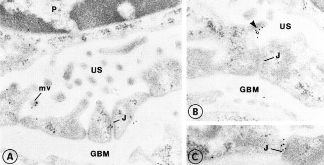
In this example of CNF, nephrin is found on the surface of flattened podocytes in association with microvilli (A-C). In occasional intercellular junctions of podocytes (J), clusters of nephrin are also found. Original magnification, ×35,000.
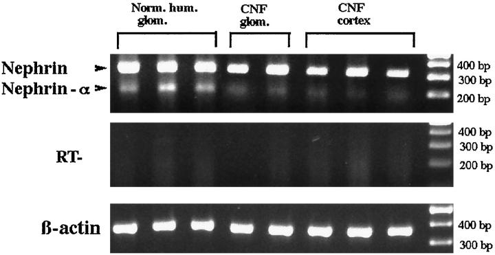
Expression pattern of nephrin after PCR analysis of mRNA from normal human glomeruli, CNF glomeruli, and CNF cortical tissue. Full-length nephrin and a splicing variant, nephrin-α, missing the whole transmembrane domain, were verified by direct sequencing as described in Materials and Methods. The control of reverse transcription shows absence of reaction product in samples without reverse transcription. β-Actin was used to verify the quality of RNA.
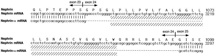
Amino acid sequence of nephrin and the respective NPHS1 nucleotides 3121–3300, including the transmembrane area (shaded) together with exon boundaries. The missing sequence of the novel nephrin-α splicing variant includes exon 24.
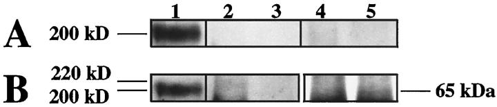
Western blotting of coimmunoprecipitations with antinephrin antibodies. Human glomerular lysate was first immunoprecipitated with anti-ZO-1 (A and B, lanes 2 and 4), antioccluding (lanes 3 and 5), or antinephrin antibodies (A and B, lane 1). Antinephrin antibodies then used for coimmunoprecipitation with ZO-1 (A, lane 2) or occludin (A, lane 3) failed to show specific coprecipitation products. The preimmune antinephrin serum (A, lanes 4 and 5) failed to precipitate any products. Control precipitation and subsequent blotting with anti-ZO-1 (B, lane 2) showed a faint band at the expected 220-kd area but no product in blots with antioccludin antibody (B, lane 3). The similar control with antioccludin immunoprecipitation showed a faint band at 65 kd (B, lane 5) but no reactivity when precipitated with anti-ZO-1 antibody (B, lane 4). Lane 1 (in A and B) shows nephrin immunoblotted with antinephrin antibodies. Subsequent blotting with the same antibody shows distinct reactivity with a 200-kd band.
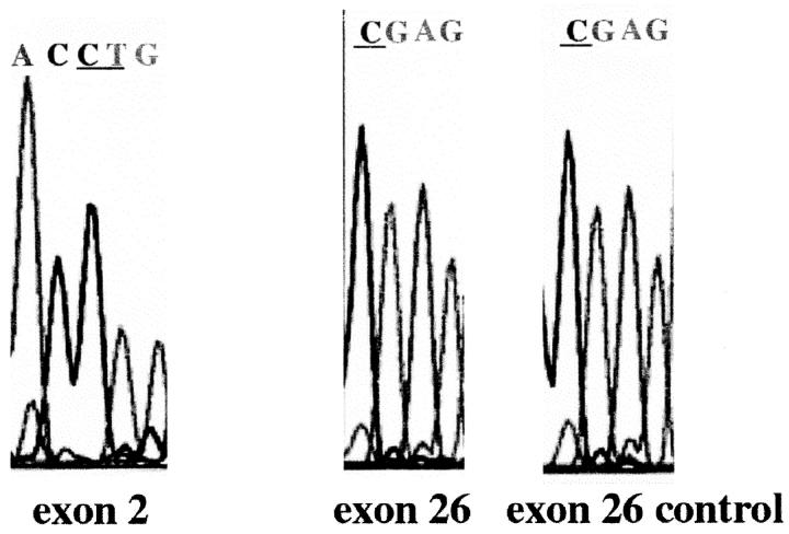
Analysis of the Finmajor (exon 2) and Finminor (exon 26) mutation sites in a patient showing positive immunoreactivity with nephrin-specific antipeptide antibodies. No mutations are found in the expected bases (underlined). For details, see Materials and Methods.
References
-
- Rennke HG: How does glomerular epithelial cell injury contribute to progressive glomerular damage. Kidney Int 1994, 45(Suppl.):S58-S63 - PubMed
-
- Kerjaschki D: Dysfunctions of cell biological mechanisms of visceral epithelial cells (podocytes) in glomerular diseases. Kidney Int 1994, 45:300-313 - PubMed
-
- Mundel P, Kriz W: Structure and function of podocytes: an update. Anat Embryol 1997, 192:385-397 - PubMed
-
- Smoyer WE, Mundel P: Regulation of podocyte structure during the development of nephrotic syndrome. J Mol Med 1998, 76:172-183 - PubMed
Publication types
MeSH terms
Substances
LinkOut - more resources
Full Text Sources
Other Literature Sources
Molecular Biology Databases
