. 1999 Nov;155(5):1749-58.
doi: 10.1016/S0002-9440(10)65490-9.

Lipopolysaccharide-activated macrophages stimulate the synthesis of collagen type I and C-fibronectin in cultured pancreatic stellate cells

Affiliations

Lipopolysaccharide-activated macrophages stimulate the synthesis of collagen type I and C-fibronectin in cultured pancreatic stellate cells

A Schmid-Kotsas et al. Am J Pathol. 1999 Nov.

Abstract

We have recently identified and characterized pancreatic stellate cells (PSC) in rats and humans (Gastroenterology 1998, 15:421-435). PSC are suggested to represent the main cellular source of extracellular matrix in chronic pancreatitis. Now we describe a paracrine stimulatory loop between human macrophages and PSC (rat and human) that results in an increased extracellular matrix synthesis. Native and transiently acidified supernatants of cultured macrophages were added to cultured PSC in the presence of 0.1% fetal calf serum. Native supernatants of lipopolysaccharide-activated macrophages stimulated the synthesis of collagen type I 1.38 +/- 0.09-fold of control and c-fibronectin 1.89 +/- 0.18-fold of control. Transiently acidified supernatants stimulated collagen type I and c-fibronectin 2.10 +/- 0.2-fold and 2.80 +/- 0.05-fold of control, respectively. Northern blot demonstrated an increased expression of the collagen-I-(alpha-1)-mRNA and fibronectin-mRNA in PSC 10 hours after addition of the acidified macrophage supernatants. Cell proliferation measured by bromodeoxyuridine incorporation was not influenced by the macrophage supernatants. Unstimulated macrophages released 1.97 pg TGFbeta1/microgram of DNA over 24 hours and lipopolysaccharide-activated macrophages released 6.61pg TGFbeta1/microgram of DNA over 24 hours. These data together with the results that, in particular, transiently acidified macrophage supernatants increased matrix synthesis, identify TGFbeta as the responsible mediator. In conclusion, our data demonstrate a paracrine stimulation of matrix synthesis of pancreatic stellate cells via TGFbeta1 released by activated macrophages. We suggest that macrophages might play a pivotal role in the development of pancreas fibrosis.


Figures
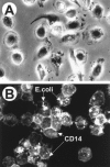
Figure 1.
Figure 1.
A: Phase contrast image of cultured macrophages. Peripheral blood mononuclear cells were isolated from buffy coats by gradient centrifugation. Cells were seeded on glass coverslips, and after 2 hours the nonadherent cells were removed by washing. Medium was changed each third day. Phase contrast micrography was performed with living cells that had been cultured for 10 days (original magnification, ×400). B: Fluorescence micrograph of cultured macrophages. CD14 immunostaining and FITC-E. coli phagocytosis of macrophages derived from peripheral blood mononuclear cells during culture for 2 weeks. Bevor fixation cells were incubated with FITC-labeled E. coli for 6 hours (original magnification, ×400).
Figure 2.
Figure 2.
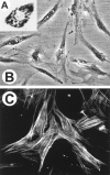
Cultured pancreatic stellate cells. A: Oil red O staining of a primary cultured rat pancreatic stellate cell. PSC were isolated by density gradient centrifugation from the pancreas of untreated male Wistar rats and seeded in a density of 4 × 10 cells/cm on glass coverslips. Eighteen hours after seeding, cells were stained using oil red O and light microscopy was performed. The cell shows a triangular shape and numerous red-stained fat droplets (original magnification, ×400). B: Phase contrast microscopy of primary cultured rat PSC. PSC were isolated and cultured as described in A. Forty-eight hours after seeding, phase contrast micoscopy was performed. Most cells are spindle-shaped and contain 6 to 20 perinuclear fat droplets (original magnification, ×200). C: Fluorescence micrograph of secondary cultured human PSC. PSC were obtained by outgrowth from small tissue blocks of fibrotic pancreas. After reaching confluency, cells were passaged and seeded on glass coverslips. Three days later, cells were fixed with acetone and indirect immunofluorescence staining was performed using anti-iso-α-sm-actin (original magnification, ×200).
Figure 3.
Figure 3.
Fluorescence micrographs showing the immunoreactivity of collagen types I and III and fibronectin in cultured human PSC stimulated with macrophage supernatants. Secondary cultured human PSC grown on glass coverslips were stimulated for 38 hours with 40 μl transiently acidified macrophage supernatant per ml medium (DMEM/Ham’s F12 with 0.1% FCS). Thereafter cultures were washed, fixed in acetone and immunostaining was performed. A-C: collagen type I; D-F: collagen type III; G-I: fibronectin. A, D, G: control (without macrophage supernatant); B, E, H: PSC stimulated with supernatant of macrophages; C, F, I: PSC stimulated with supernatant of LPS-activated macrophages. Intensive staining patterns were observed in cultures receiving supernatants of LPS activated macrophages. Collagens were detected predominantly intracellularly. Fibronectin was located predominantly extracellularly as fibrils, varying in staining intensity and in density. There was no significant difference in staining patterns between untreated cells (A, D, G) and PSC incubated with conditioned media of unstimulated macrophages (B, E, H). Original magnification, ×400.
Figure 4.
Figure 4.
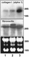
Northern blot hybridization of collagen α1(I), fibronectin, and the 18S ribosomal RNA. Transiently acidified macrophage supernatants were added to secondary cultured human PSC. Ten hours later, cultures were stopped and RNA was isolated. RNA (30 μg/lane) was separated by agarose gel electrophoresis and transferred to Hybond-N membranes. Ethidium bromide staining of the agarose gels was used to verify equal loading and blotting of total RNA. Hybridizations were performed by standard procedures using 0.5 × 10 cpm/ml labeled cDNA. Exposure to X-ray films was done at −70°C for 7 to 10 days. Lane 1: RNA isolated from unstimulated PSC (control); Lane 2: RNA isolated from PSC stimulated with supernatant of macrophages; Lane 3: RNA isolated from PSC stimulated with supernatant of LPS-activated macrophages.
Figure 5.
Figure 5.
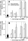
Effect of macrophage supernatants on fibronectin synthesis of cultured pancreatic stellate cells. Native and acidified macrophage supernatants (40 and 160 μl/ml medium) were added to secondary cultured PSC (A, human PSC; B, rat PSC). After 24 hours, cultures were stopped and fibronectin was measured in PSC supernatants; DNA was measured in the cell layer. Values are expressed as means ± SD of three independent experiments, each condition performed in triplicate culture wells. *P < 0.05.
Figure 6.
Figure 6.
Effect of macrophage supernatants on the synthesis of collagen type I in cultured human pancreatic stellate cells. Native and acidified macrophage supernatants (40 and 160 μl/ml medium) were added to pancreatic stellate cells cultured in DMEM/Hams F12 in the presence of 0.1% FCS, ascorbic acid (100 μg/ml) and β-aminopropionitrile (100 μg/ml). After 24 hours cultures were stopped and collagen type I was measured in PSC supernatants; DNA was measured in the cell layer. Values are expressed as means ± SD of three independent experiments, each condition performed in triplicate culture wells. *P < 0.05.

References

    1. Sarles H, Bernard JP, Johnson C: Pathogenesis and epidemiology of chronic pancreatitis. Ann Rev Med 1989, 40:453-468 - PubMed
    1. Tsukamoto H, Towner SJ, Yu GSM, French SW: Potentiation of ethanol-induced pancreatic injury by dietary fat. Am J Pathol 1988, 131:246-257 - PMC - PubMed
    1. Morohoshi T, Kanda M: Periacinar fibroblastoid cell: its action on early stage of alcoholic pancreatitis. J Biliary Tract Pancreas 1985, 6:1205-1211(in Japanese)
    1. Di Magno EP, Layer P, Clain JE: Chronic pancreatitis. 21st ed. Go VWL Di Magno EP Gardner JD eds. The Pancreas, 1993, :pp 655-706 Raven Press, New York
    1. Elsässer HP, Adler G, Kern HF: Fibroblast structure and function during regeneration from hormone-induced acute pancreatitis in the rat. Pancreas 1989, 4:169-178 - PubMed

Publication types

LinkOut - more resources