. 1999;8(2):115-27.

Constitutive and inducible in vivo protein-DNA interactions at the tumor necrosis factor-alpha promoter in primary human T lymphocytes

Affiliations

Constitutive and inducible in vivo protein-DNA interactions at the tumor necrosis factor-alpha promoter in primary human T lymphocytes

C Becker et al. Gene Expr. 1999.

Abstract

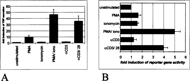
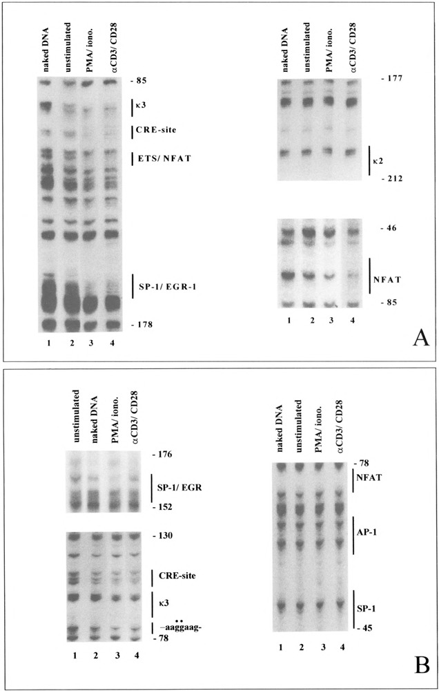
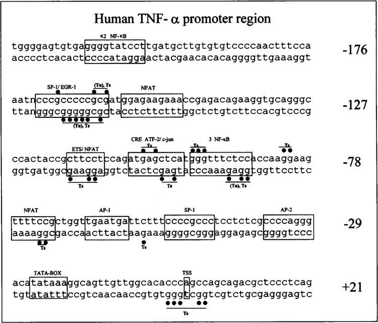
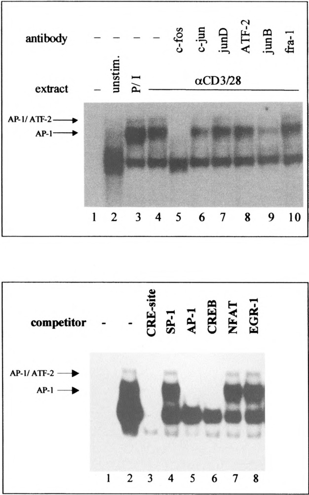
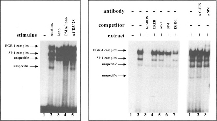
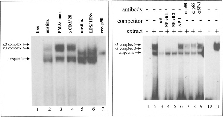
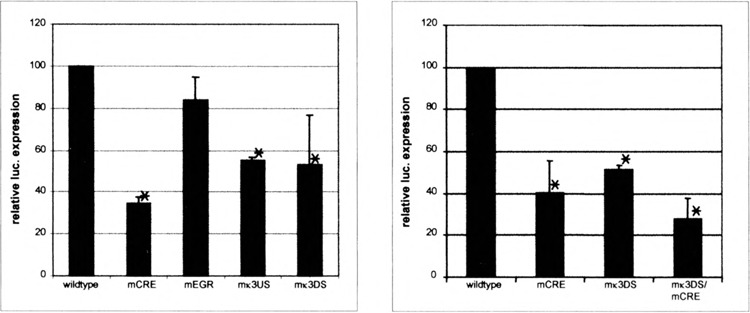
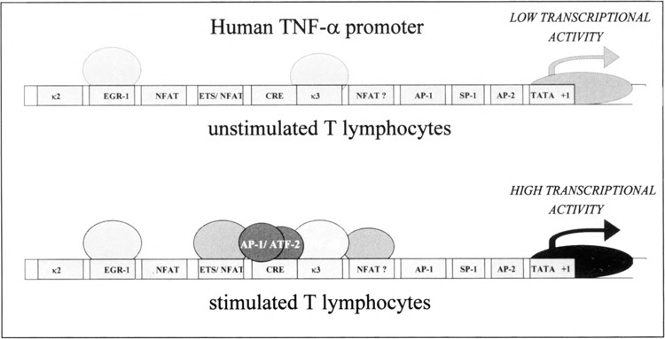
Tumor necrosis factor-alpha (TNF-alpha) is a key cytokine of lymphocytes with major regulatory functions in immunomodulation, chronic inflammation, and septic shock. However, only limited information on TNF promoter regulation in vivo in primary lymphocytes is available. To determine and compare protein-DNA interactions at the native TNF locus in primary lymphocytes, we analyzed the human TNF-alpha promoter by ligation-mediated polymerase chain reaction (LM-PCR) techniques. Accordingly, primary CD4+ T lymphocytes from peripheral blood were cultured in the presence of various stimuli and analyzed by LM-PCR. Inducible in vivo protein-DNA interactions at the TNF promoter were detected between -120 and -70 bp of the human TNF promoter relative to the transcriptional start site. This area includes binding sites for transcription factors such as ETS-1, NFAT, ATF-2/c-jun, SP-1/Egr-1, and NF-kappaB. In contrast, no protein-DNA interactions were observed at various binding sites with reported regulatory function in tumor cell lines such as the k2 element, the NFAT site at -160, the AP1 site at -50, and the SP1 site at -65. Additional mutagenesis and transfection studies demonstrated that NF-kappaB and CREB/AP-1 are important regulators of inducible TNF promoter activity in primary human T lymphocytes. These results provide novel insights into the complex regulation of TNF gene transcription in primary T lymphocytes in vivo by constitutive and inducible protein-DNA interactions that appear to be at least partially different compared to previously characterized tumor cell lines.


Figures

FIG.1
FIG.1
TNF is inducible in freshly isolated human T lymphocytes. (A) TNF protein production by stimulated and unstimulated primary T lymphocytes. Primary T lymphocytes were stimulated with PMA, ionomycin (iono), or antibodies to CD3/CD28, as specified in Materials and Methods. Cell-free supernatant was collected after 48 h and cytokine concentration was determined by ELISA. Data were normalized to the values from unstimulated control samples and represent mean values ± SD of three independent experiments. (B) Activity of TNF promoter constructs in primary CD4+ T cells. Primary CD4+ T lymphocytes were transfected with the pXPl-TNF-Luc vector containing the human TNF promoter upstream of a luciferase reporter gene (see Materials and Methods). Cells were stimulated with indicated stimuli 24 h after transfection and luciferase activity was assessed after an additional 18 h. Data represent mean values ± SD of three independent experiments and are expressed as fold induction of each stimulation condition compared to the unstimulated control.
FIG. 2
FIG. 2
In vivo protein-DNA interactions at the TNF promoter in primary T lymphocytes. CD4+ T lymphocytes were stimulated for 4 h prior to DMS treatment. After isolation of methylated DNA the DNA was subjected to the LM-PCR method and analyzed by denaturing polyacrylamide electrophoresis. Footprinting ladders on the coding (A) and noncoding (B) strands were compared to DMS reactivity of naked DNA that was treated with DMS in vitro. The location of previously described binding sites of transcription factors is indicated.
FIG. 3
FIG. 3
Summary of in vivo protein-DNA interactions in the 5′ flanking region of the TNF gene in unstimulated (Tu) and PMA plus ionomycin-stimulated (Ts) CD4+ T lymphocytes. Circles indicate protected bases. The location of previously characterized binding sites for transcription factors in the TNF promoter region is indicated.
FIG. 4
FIG. 4
Upper panel: Activation-induced changes of nuclear proteins binding to the CRE site of the human TNF promoter in primary CD4+ T lymphocytes. Nuclear proteins were extracted from primary cells 1 h after activation with indicated stimuli. Two in-ducible specific complexes were seen using extracts from primary T lymphocytes. For supershift assays antibodies were added to the binding reaction as indicated. Fower panel: Competition assays at the CRE site using nuclear extracts from PMA plus ionomycin-stimulated CD4+ T lymphocytes. Competitor DNA was added to the binding reaction as indicated.
FIG. 5
FIG. 5
Left panel: Activation-induced changes of nuclear proteins binding to the GC-box of the human TNF promoter in primary CD4+ T lymphocytes. Two specific complexes were observed in the gel retardation studies as indicated. Right panel: Competition and supershift analyses at the GC-box using nuclear extracts from PMA plus ionomycin-stimulated CD4+ T lymphocytes. Antibodies (2 μg) or competitor DNA were added to the binding reaction as indicated. A supershift of the lower complex was observed after addition of antibodies to SP-1.
FIG. 6
FIG. 6
Left panel: Binding analysis at the k3 site using nuclear extracts from unstimulated and stimulated T lymphocytes (lanes 1–4) and CD 14+ monocytes (lanes 5–6). An inducible specific double complex (upper complex: 1; lower complex: 2) was found upon stimulation with PMA/ionomycin or anti-CD3/CD28 in T cells. Two complexes at the k3 site were found in monocytes: an inducible complex (lane 6) was observed that comigrated with complex 2 in T cells. Furthermore, a slower migrating constitutive complex was seen in CD 14+ monocytes that comigrated with recombinant p50. Right panel: Competition and supershift assays at the k3 site using nuclear extracts from stimulated T lymphocytes. The specific complexes could be cross-competed with two different NF-κB consensus oligonucleotides and were reduced in intensity after addition of antibodies to the p50 subunit of NF-κB. Antibodies to p65 caused a reduced intensity of the lower complex, suggesting the presence of p50/p65 in complex 2.
FIG. 7
FIG. 7
Transfection analysis of TNF promoter constructs carrying single or double mutations of the CRE/Egr-1 sites and the proximal or distal parts of the k3 site, respectively. Wild-type and mutagenized luciferase reporter gene constructs were transfected into PMA plus ionomycin-stimulated primary human CD4 ± T cells. Data represent mean values ± SD of four (left panel) and three (right panel) independent experiments after normalization for transfection efficiency. Luciferase activity is reported as percentage compared to T cells transfected with the wild-type construct (100%).
FIG. 8
FIG. 8
Schematic model of protein-DNA interactions at the TNF promoter in vivo in unstimulated and PMA plus ionomycin-stimulated primary human CD4+ T lymphocytes. Dotted lines indicate weak protein-DNA interactions in vivo.

Similar articles

Cited by

References

    1. Barbulescu K.; Becker C.; Schlaak J. F.; Schmitt E.; Meyer-zum-Büschenfelde K.-H.; Neurath M. F. IL-12 and IL-18 differentially activate the human IFN-gamma promoter in primary CD4+ T lymphocytes. J. Immunol. 160:3642–3649; 1998. - PubMed
    1. Barbulescu K.; Meyer-zum-Büschenfelde K.-H.; Neurath M. F. Constitutive and inducible protein/ DNA interactions of the interferon-gamma promoter in vivo in CD45RA and CD45R0 T helper subsets. Eur. J. Immunol. 27:1098–1108; 1997. - PubMed
    1. Campbell I. L.; Iscaro A.; Harrison K. IFN-gamma and tumor necrosis factor-alpha. Cytotoxicity to murine islets of Langerhans. J. Immunol. 141:2325–2329; 1988. - PubMed
    1. Collart M. A.; Belin D.; Vassalli J. D.; de-Kossodo K. S.; Vassalli P. Gamma interferon enhances macrophage transcription of the tumor necrosis factor/cachectin, interleukin 1, and urokinase genes, which are controlled by short-lived repressors. J. Exp. Med. 164:2113–2118; 1986. - PMC - PubMed
    1. Di G. N.; Visvanathan K.; Lloyd A.; Wakefield D. Expression of TNF-alpha by human plasma cell in chronic inflammation. J. Leukoc. Biol. 61:667–678; 1997. - PubMed

Publication types