

Neural correlates of memories of childhood sexual abuse in women with and without posttraumatic stress disorder
- PMID: 10553744
- PMCID: PMC3233772
- DOI: 10.1176/ajp.156.11.1787
Neural correlates of memories of childhood sexual abuse in women with and without posttraumatic stress disorder
Abstract
Objective: Childhood sexual abuse is very common in our society, but little is known about the long-term effects of abuse on brain function. The purpose of this study was to measure neural correlates of memories of childhood abuse in sexually abused women with and without the diagnosis of posttraumatic stress disorder (PTSD).
Method: Twenty-two women with a history of childhood sexual abuse underwent injection of [15O]H2O, followed by positron emission tomography imaging of the brain while they listened to neutral and traumatic (personalized childhood sexual abuse events) scripts. Brain blood flow during exposure to traumatic and neutral scripts was compared for sexually abused women with and without PTSD.
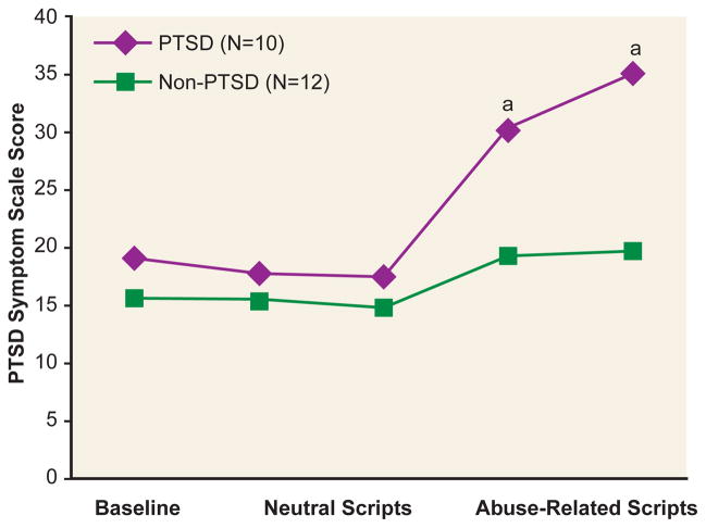
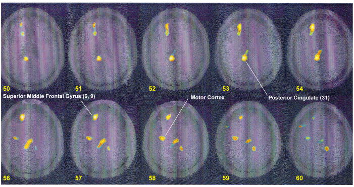
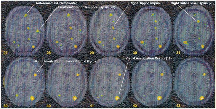
Results: Memories of childhood sexual abuse were associated with greater increases in blood flow in portions of anterior prefrontal cortex (superior and middle frontal gyri-areas 6 and 9), posterior cingulate (area 31), and motor cortex in sexually abused women with PTSD than in sexually abused women without PTSD. Abuse memories were associated with alterations in blood flow in medial prefrontal cortex, with decreased blood flow in subcallosal gyrus (area 25), and a failure of activation in anterior cingulate (area 32). There was also decreased blood flow in right hippocampus, fusiform/inferior temporal gyrus, supramarginal gyrus, and visual association cortex in women with PTSD relative to women without PTSD.
Conclusions: These findings implicate dysfunction of medial prefrontal cortex (subcallosal gyrus and anterior cingulate), hippocampus, and visual association cortex in pathological memories of childhood abuse in women with PTSD. Increased activation in posterior cingulate and motor cortex was seen in women with PTSD. Dysfunction in these brain areas may underlie PTSD symptoms provoked by traumatic reminders in subjects with PTSD.
Figures
References
-
- McCauley J, Kern DE, Kolodner K, Dill L, Schroeder AF, DeChant HK, Ryden J, Derogatis LR, Bass EG. Clinical characteristics of women with a history of childhood abuse: unhealed wounds. JAMA. 1997;277:1362–1368. - PubMed
-
- Kessler RC, Sonnega A, Bromet E, Hughes M, Nelson CB. Posttraumatic stress disorder in the national comorbidity survey. Arch Gen Psychiatry. 1995;52:1048–1060. - PubMed
-
- Pitman RK. Posttraumatic stress disorder, hormones, and memory (editorial) Biol Psychiatry. 1989;26:221–223. - PubMed
-
- Bremner JD, Krystal JH, Southwick SM, Charney DS. Functional neuroanatomical correlates of the effects of stress on memory. J Trauma Stress. 1995;8:527–554. - PubMed
-
- Roth RH, Tam SY, Ida Y, Yang JX, Deutch AY. Stress and the mesocorticolimbic dopamine systems. Ann NY Acad Sci. 1988;537:138–147. - PubMed
Publication types
MeSH terms
Grants and funding
LinkOut - more resources
Full Text Sources
Other Literature Sources
Medical
