. 1999 Nov 4;9(21):1267-70.
doi: 10.1016/s0960-9822(99)80510-7.

Postsynaptic expression of tetanus toxin light chain blocks synaptogenesis in Drosophila

Affiliations

Postsynaptic expression of tetanus toxin light chain blocks synaptogenesis in Drosophila

R A Baines et al. Curr Biol. .

Abstract

During the development of the nervous system embryonic neurons are incorporated into neural networks that underlie behaviour. For example, during embryogenesis in Drosophila, motor neurons in every body segment are wired into the circuitry that drives the simple peristaltic locomotion of the larva. Very little is known about the way in which the necessary central synapses are formed in such a network or how their properties are controlled. One possibility is that presynaptic and postsynaptic elements form relatively independently of each other. Alternatively, there might be an interaction between presynaptic and postsynaptic neurons that allows for adjustment and plasticity in the embryonic network. Here we have addressed this issue by analysing the role of synaptic transmission in the formation of synaptic inputs onto identified motorneurons as the locomotor circuitry is assembled in the Drosophila embryo. We targeted the expression of tetanus toxin light chain (TeTxLC) to single identified neurons using the GAL4 system. TeTxLC prevents the evoked release of neurotransmitter by enzymatically cleaving the synaptic-vesicle-associated protein neuronal-Synaptobrevin (n-Syb) [1]. Unexpectedly, we found that the cells that expressed TeTxLC, which were themselves incapable of evoked release, showed a dramatic reduction in synaptic input. We detected this reduction both electrophysiologically and ultrastructurally.


Figures

Figure 1
Figure 1
Identification of the aCC and RP2 motorneurons. (a) The aCC motorneuron has a dendritic process (arrowhead) extending from the cell body in addition to its axon (arrow). (b) The RP2 motorneuron has only a single axonal projection (arrow). The patch electrode (asterisk) is shown in place in both images. The scale bars represent 10 µm.
Figure 2
Figure 2
TeTxLC expression reduces synaptic activity. (a) Recordings, under voltage clamp (Vh = −60 mV), from an aCC (upper trace) and RP2 (lower trace) neuron show the presence of discrete epscs. (b) Recordings from aCC and RP2 neurons also reveal the presence of relatively slow and large sustained inward currents which often support a burst of action potentials (arrow). (c,d) The proportion of aCC and RP2 neurons that exhibited either (c) epscs or (d) sustained inward currents in the control parental lines (GAL4 and UAS) and when TeTxLC was expressed throughout the CNS. The white bars represent neurons in which these respective currents were observed (labelled +) and the grey bars represent neurons in which currents were not seen (labelled −). The given values represent the frequency per min of observed currents ± standard error (SE).
Figure 3
Figure 3
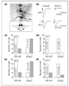
Selective expression of TeTxLC in aCC neurons deprives them of synaptic input. (a) Expression pattern of RRC-GAL4 visualised by anti-β-galactosidase immunostaining in three abdominal segments of a late stage 17 embryo (20 h AEL). Two strongly stained aCC neurons are present in all three segments (arrows), whereas staining of RP2 is more variable (arrowheads) and pCC staining is weak (open arrowhead). Only one pCC cell is visible in the three segments shown. The scale bar represents 10 µm. The inset shows a diagrammatic representation of the relative positions of aCC, pCC and RP2 neurons in a single abdominal segment in a late stage 17 embryo. Neurons are shaded to reflect their levels of RRC-GAL4 expression. (b) Electrical stimulation of the axon of an aCC neuron that is expressing TeTxLC under the control of RRC-GAL4 failed to evoke an ejc in its target muscle DA1. Immunostaining of the terminals of aCC using an anti-Fasciclin II antibody revealed no obvious morphological abnormalities (data not shown). Synaptic communication between other motorneurons and their target muscles was not affected (RP3 and muscle VL3 are shown). The RRC-GAL4 line was used as control. (c) Selective expression of TeTxLC results in a significant reduction in the number of aCC neurons that exhibit epscs (white bars, +) compared with expression of inactive toxin (TNT-VIF). (d) The proportion of pCC neurons that exhibit epscs (white bars, +), and their frequency, does not differ significantly in embryos expressing either TeTxLC or TNT-VIF. (e) Expression of TeTxLC similarly results in a significant decrease in the number of aCC neurons that show sustained inward currents compared to TNT-VIF (white bars, +). (f) Expression of TeTxLC does not affect the appearance or frequency of sustained currents in pCC. In each case, the white bars represent those neurons in which these respective currents were observed (+) and the grey bars represent those neurons in which currents were not seen (−). In (c–f) the given values represent the frequency per min of observed currents ± SE.
Figure 4
Figure 4
Identification of synaptic input to aCC. Electron micrographs showing accumulations of clear synaptic vesicles (arrowheads) immediately adjacent to labelled profiles of aCC neurons (asterisks) in (a) a TNT-VIF and (b) a TeTxLC background (driven by RRC-GAL4). The scale bars represent 200 nm. Potential sites of synaptic input were identified by the accumulation of vesicles, some of which are docked to the presynaptic membrane, immediately adjacent to labelled profiles of aCC. At this stage of development, synaptic elements such as T-bars are not present although increased electron density at the presynaptic membrane is visible in some instances (a). (c) Although similar in appearance, the frequency of putative presynaptic elements adjacent to aCC neurons expressing TeTxLC is significantly reduced (p ≤ 0.01, Chi squared test) compared to control (TNT-VIF).

References

    1. Sweeney ST, Broadie K, Keane J, Niemann H, O’Kane J. Targeted expression of tetanus toxin light chain in Drosophila specifically eliminates synaptic transmission and causes behavioral defects. Neuron. 1995;14:341–351. - PubMed
    1. Baines RA, Bate M. Electrophysiological development of central neurons in the Drosophila embryo. J Neurosci. 1998;18:4673–4683. - PMC - PubMed
    1. Landgraf M, Bossing T, Technau GM, Bate M. The origin, location, and projections of the embryonic abdominal motorneurons of Drosophila. J Neurosci. 1997;15:9642–9655. - PMC - PubMed
    1. Salvaterra PM, McCaman RE. Choline acetyltransferase and acetycholine levels in Drosophila melanogaster: a study using two temperature-sensitive mutants. J Neurosci. 1985;5:903–910. - PMC - PubMed
    1. Mlodzik M, Baker NE, Rubin GM. Isolation and expression of scabrous, a gene regulating neurogenesis in Drosophila. Genes Dev. 1990;4:1848–1861. - PubMed

Publication types

LinkOut - more resources