. 1999 Nov 1;13(21):2787-800.
doi: 10.1101/gad.13.21.2787.

Progressive impairment of developing neuroendocrine cell lineages in the hypothalamus of mice lacking the Orthopedia gene

Affiliations

Progressive impairment of developing neuroendocrine cell lineages in the hypothalamus of mice lacking the Orthopedia gene

D Acampora et al. Genes Dev. .

Abstract

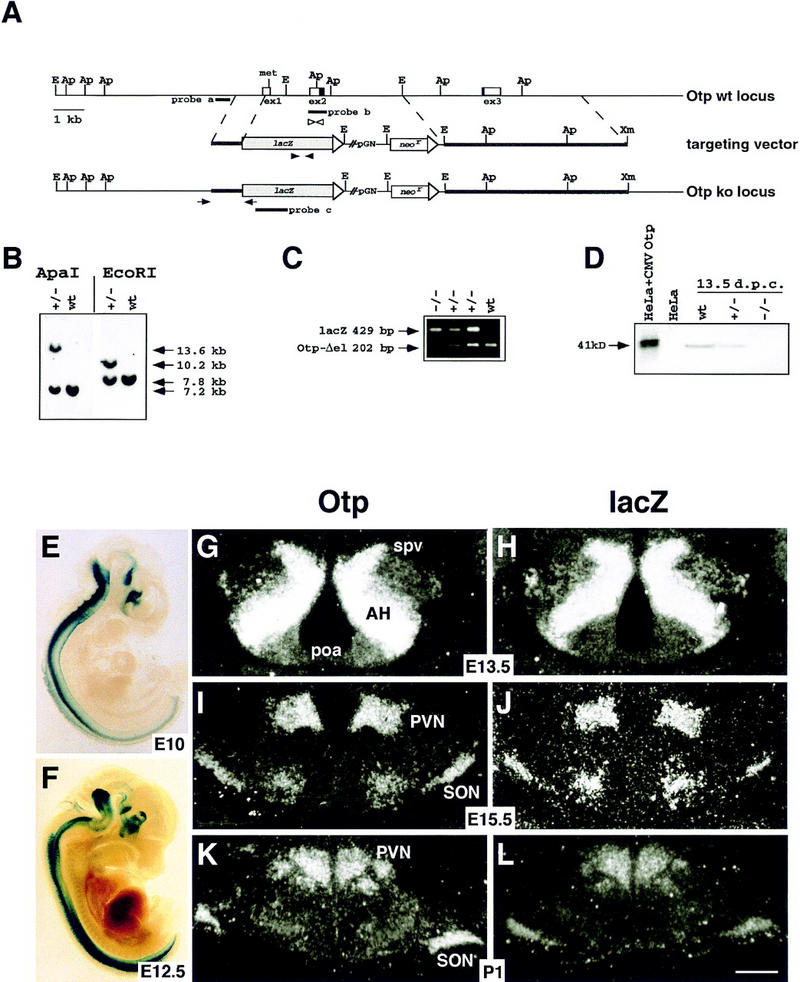
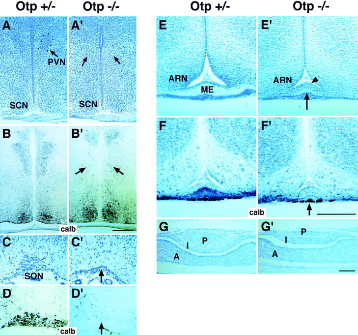
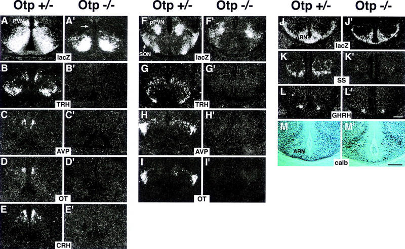
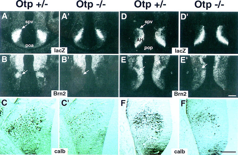
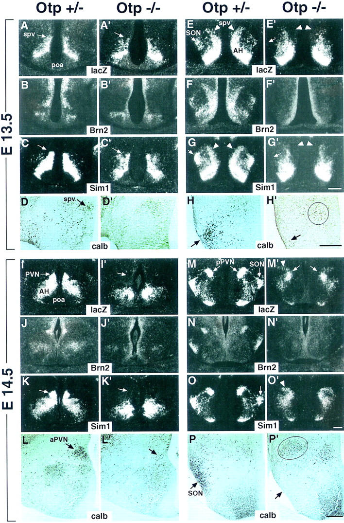
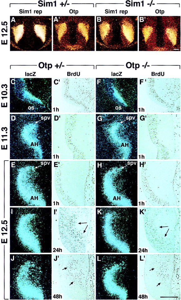
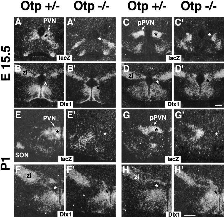
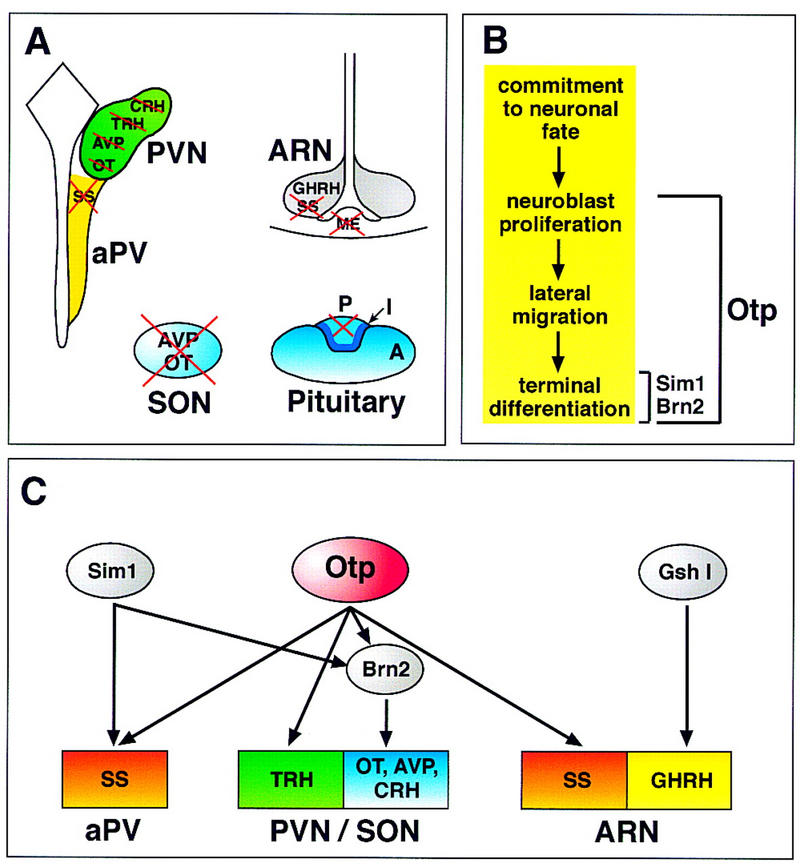
Development of the neuroendocrine hypothalamus is characterized by a precise series of morphogenetic milestones culminating in terminal differentiation of neurosecretory cell lineages. The homeobox-containing gene Orthopedia (Otp) is expressed in neurons giving rise to the paraventricular (PVN), supraoptic (SON), anterior periventricular (aPV), and arcuate (ARN) nuclei throughout their development. Homozygous Otp(-/-) mice die soon after birth and display progressive impairment of crucial neuroendocrine developmental events such as reduced cell proliferation, abnormal cell migration, and failure in terminal differentiation of the parvocellular and magnocellular neurons of the aPV, PVN, SON, and ARN. Moreover, our data provide evidence that Otp and Sim1, a bHLH-PAS transcription factor that directs terminal differentiation of the PVN, SON, and aPV, act in parallel and are both required to maintain Brn2 expression which, in turn, is required for neuronal cell lineages secreting oxytocin (OT), arginine vasopressin (AVP), and corticotropin-releasing hormone (CRH).


Figures

Figure 1
Figure 1
Targeted replacement of Otp with the lacZ gene. (A) Schematic representation of the wild-type Otp allele, targeting vector, and recombined allele. E, Ap and Xm stand for EcoRI, ApaI, and XmnI. The locations of probes a, b, and c used for Southern blot analyses (probes a and c) and in situ hybridizations (probes b and c) are indicated. The arrows in the third line indicate the position of the primers used to identify homologous recombinant clones. (B) Southern blot analysis of one targeted ES cell line and wild-type HM-1 ES cells showing expected hybridization pattern of ApaI and EcoRI-digested genomic DNA samples with probe a (A). Only the 13.6-kb band with ApaI and the 10.2-kb band with EcoRI were detected with the lacZ internal probe c (data not shown). (C) Genotyping of littermates from the intercross of heterozygotes by a PCR reaction amplifying fragments specific for the deleted region of Otp (OtpΔel) (202 bp) and lacZ (429 bp), with the primers indicated as open and filled arrowheads, respectively. (D) Western blot analysis of the OTP protein in transfected HeLa cells and embryonic extracts. (EL) β-gal staining of Otp+/− embryos at E10 (E) and E12.5 (F) and in situ hybridization on adjacent sections of Otp+/− brains at E13.5 (G,H), E15.5 (I,J), and P1 (K,L) with allele-specific probes (probes b and c in A) revealing no difference between the wild-type and the targeted locus. (spv) Supraoptic/paraventricular area; (AH) anterior hypothalamus; (PVN) paraventricular nucleus; (poa) anterior preoptic area; (SON) supraoptic nucleus. For sections in GL, scale bar corresponds to 50 μm (L).
Figure 2
Figure 2
Abnormalities in the hypothalamus and pituitary of Otp−/− mice. (AF') Nissl staining (A,A',C,C',E,E') and calb-positive cells (B,B',D,D',F,F') in sections through the PVN (A,B'), SON (C,D'), and ME (E,F') of Otp+/− (AF) and Otp−/− (A'F') newborns. Arrows in A'D' indicate the lack of the PVN (A') and SON (C') confirmed by the absence of calb-positive cells (B',D'). The arrows in E' and F' point to the heavy reduction in thickness of the ME (E') that results poorly stained with α-calb antibody (F'). Note that the neuroepithelium adjacent to the presumptive ME results in increased thickness (arrowhead in E'). (G,G') As compared with Otp+/− (G), the posterior lobe of the pituitary gland is strongly hypoplastic in Otp−/− mice (G'). Abbreviations as in the previous figure; (ME) median eminence; (SCN) suprachiamastic nucleus; (ARN) arcuate nucleus; (A) I, P anterior, intermediate, and posterior lobes of the pituitary. Scale bars, 50 μm. Scale bar in B' refers to A and B'; scale bar in F' to CF', and scale bar in G' to GG'.
Figure 3
Figure 3
Abnormal lacZ expression and lack of neuroendocrine hormones in Otp−/− brain. (AC') Coronal sections of Otp+/− (AC) and Otp−/− (A'C') brains at P1 showing that lacZ is expressed in Otp+/− brain all along the PVN (AC), aPV (A), AH (A), and SON (B), whereas in Otp−/−, brain, lacZ transcripts disappear from the presumptive areas corresponding to the PVN (A'C'), aPV (A'), AH (A'), and SON (B') and are detected in a misplaced domain that normally never expresses Otp (A'C'). (D,D') In sections through the ARN of Otp+/− (D) and Otp−/− (D') brains, lacZ is expressed in the ARN, in a narrow domain adjacent to the ARN (arrowheads) and in a region possibly including the premammillary nucleus. Note the remarkable reduction in lacZ expression in Otp−/− brains. (EK') In Otp+/− brain (EK), TRH (E) is detected in the PVN and in scattered neurons, CRH (F) in the PVN, AVP (G) in the PVN, SON, and SCN, OT (H) in the PVN and SON, SS (I,J) in numerous areas and among them in the aPV (I) and ARN (J) and GHRH in the ARN (K), whereas in Otp−/− brain (E'K'), TRH (E'), CRH (F'), AVP (G'), and OT (H') are undetectable in the PVN and SON, SS is not transcribed in the aPV (I') and ARN (J'), whereas GHRH (K') appears unaffected. Also note that in Otp−/− mice, TRH expression totally disappears in the hypothalamus; AVP expression is retained in the SCN (G'), and SS-positive neurons are not identified in the area surrounding the ARN (J'). Abbreviations as in the previous figures; (aPV) anterior periventricular nucleus; (pMA) premammillary nucleus. Scale bar, 50 μm.
Figure 4
Figure 4
Failure in transcriptional activation of neuroendocrine hormones in Otp−/− embryos. (AL') Frontal sections through the hypothalamus of E14.5 Otp+/− (AL) and Otp−/− (A'L') embryos showing that at the level of the poa area (AE') lacZ transcripts disappear from the presumptive PVN primordium (arrow in A') and TRH (B'), AVP (C'), OT (D'), and CRH (E') hormone transcripts are not detected; at the level of the pPVN (FI') lacZ is abnormally transcribed (F') and no expression appears for the TRH (G'), AVP (H'), and OT (I'); and at the level of the ARN (JL') no macroscopic abnormalities are detected in the distribution of lacZ (J') and GHRH (L') transcripts, whereas SS-positive neurons disappear (K'). (M,M') As compared with Otp+/− (M), a considerable reduction in the number of calb-positive cells is observed in the ARN and surrounding areas of Otp−/− embryos (M'). Abbreviations as the previous figures. Scale bars, 50 μm. Scale bar in L' refers to dark fields, and in M' to M, and M'.
Figure 5
Figure 5
Abnormalities in Otp−/− embryos at E12.5. (AF') Otp+/− (AF) and Otp−/− (A'F') embryos hybridized with the indicated lacZ (A,A',D,D') and Brn2 (B,B',E,E') probes or processed for detecting calb-positive cells (C,C',F,F') in sections adjacent to those hybridized with radioactive probes and photographed at a higher magnification. Note that Otp−/− embryos show narrowed expression domains of lacZ (A',D'); lack of Brn2 transcripts in the spv (arrow in B',E'); and, finally, a remarkable reduction in the number of calb-positive cells (C',F'). Abbreviations as in the previous figures; in C,C',F,F' the third ventricle is at right. Scale bars, 50 μm. Scale bar in E' refers to dark fields and in F' to bright fields.
Figure 6
Figure 6
Abnormalities in Otp−/− embryos at E13.5 and E14.5. (AP') Adjacent frontal sections of Otp+/− (AP) and Otp−/− (A'P') embryos at E13.5 (AH') and E14.5 (IP') are hybridized with the indicated lacZ, Brn2, and Sim1 or stained for detecting calb-positive cells. At E13.5 in sections through the poa area of Otp−/− embryos, lacZ (A') and Sim1 (C') transcripts are disorganized and poorly condensed in the spv and in sections through the pop area; the lacZ and Sim1 expression disappears from the presumptive SON (cf. arrows in E,G with arrows in E',G') and is heavily affected in the spv region (cf. arrowheads in E,G with arrowheads in E',G'). At E14.5 in sections through the poa area, lacZ and Sim1 expression is not detected in the PVN primordium (arrow in I',K') and at the level of pPVN, a fraction of migrating lacZ- (M') and Sim1- (O') positive cells is still identified. In E13.5 and E14.5 Otp−/− embryos, only sporadic calb-positive cells are identified in the spv/PVN primordium (cf. D with D' and arrow in L with arrow in L') and in the presumptive SON (cf. arrow in H,P with arrow in H',P', respectively). Note that the circled group of calb-positive cells (P') that is coincident with the lacZ and Sim1 expression (arrowhead in M',O') may, possibly, correspond to residual cells that would be committed to migrate and form the SON. Finally, Brn2 expression in the spv, PVN, SON, and pPVN of Otp+/− embryos (B,F,J,N) is undetectable in Otp−/− mutants (B',F',J',N'). Note that calb-staining sections (D,D', H,H',L,L',P,P') are adjacent to those hybridized with radioactive probes, but are photographed at a higher magnification. Abbreviations as in the previous figures and in L,L',P,P', the third ventricle is at right. Scale bars, 50 μm. Scale bar in G' refers to AC' and EG'; scale bar in O' to IK' and MO'; scale bar in H' to D,D',H,H'; scale bar in P' to L,L',P,P'.
Figure 7
Figure 7
Otp expression in Sim1−/− mutants and cell proliferation in Otp−/− embryos. (AB') Otp expression (A',B') was compared with that of the Sim1 reporter (Sim1 rep) (A,B) in E12.5 Sim1+/− (A,A') and Sim1−/− (B,B') embryos, showing no obvious difference between the two genotypes, thus indicating that Sim1 was not required for Otp expression. (CH') lacZ expression (CH) and BrdU-positive cells (C'H') labeled 1 hr after the injection were compared in Otp+/− and Otp−/− embryos at E10.3 (C,C',F,F'), E11.3 (D,D',G,G'), and E12.5 (E,E',H,H') showing no obvious difference between the two genotypes. Note that lacZ was expressed in the proliferating neuroepithelium only in the portion close to the mantle zone. (IL') At E12.5, comparison between lacZ expression (IL) and BrdU positive cells (I'L') detected 24 (I',K') and 48 hr (J',L') after a single injection of BrdU reveals that, as compared with Otp+/− (I,I',J,J'), in Otp−/− (K,K',L,L') embryos, the lacZ expression domain was thinner (K,L) and the number of BrdU-positive cells (K',L') remarkably reduced. Abbreviations as in the previous figures; (os) optic stalk. Arrows in I', J', K', and L' point to BrdU-positive cells that are diminished in Otp−/− embryos. Scale bars, 50 μm. Scale bar in B' refers to AB' and in L' to CL'.
Figure 8
Figure 8
Patterning abnormalities in Otp−/− mutants. (AH') Adjacent frontal sections through the hypothalamus of Otp+/− (AH) and Otp−/− (A'H') at E15.5 (AD') and P1 (EH') hybridized with lacZ and Dlx1 showing that in the PVN of Otp+/− brain, lacZ and Dlx1 show complementary expression (cf. black asterisk in A,C,E,G with white asterisk in B,D,F,H), where as in the area corresponding to the presumptive PVN of Otp−/− mutants, the lacZ and Dlx1 complementary expression was inverted (cf. white asterisk in A',C',E',G' with black asterisk in B',D',F',H'). Abbreviations as in previous figures; (zi) zona incerta. Scale bars, 50 μm. Scale bar in D' refers to E15.5 and in H' to P1 brains.
Figure 9
Figure 9
Schematic summary illustrating neuroendocrine impairments, abnormal developmental processes, and genetic interactions deduced from the analysis of Otp−/− mice. (A) In wild-type mice magnocellular and parvocellular neurons of the PVN, SON, aPV, and ARN secrete OT, AVP, CRH, TRH, SS, and GHRH neuropeptides. Magnocellular neurons located in the PVN and SON project their axons to the posterior lobe of the pituitary, whereas parvocellular neurons of the PVN, aPV, and ARN project to the ME. In Otp−/− mice, failure in terminal differentiation results in morphological disruption of PVN and SON, lack of CRH, TRH, AVP, OT, and SS neuropeptides expression, and impaired axonal outgrowth to the ME and posterior pituitary. (B) Developmental milestones of neuroendocrine hypothalamus are represented by the early commitment to the neuronal fate, neuroblast proliferation, lateral migration of postmitotic neurons to the area of nuclei formation, and terminal differentiation including neuropeptide expression, axonal outgrowth, and maintenance of cell viability. In Brn2−/− and Sim1−/− mice, terminal differentiation of cell lineages secreting CRH, AVP, OT (for Brn2), and CRH, AVP, OT, TRH, and SS (for Sim1) is disrupted. In Otp−/− mice, beside the failure in terminal differentiation of CRH, AVP, OT, TRH, and SS cell lineages, there is also an impairment in both neuroblast proliferation, and their subsequent migration, indicating that Otp is an important requirement of most of the crucial steps necessary for proper development of the neuroendocrine hypothalamus. (C) Otp is required from E12 onward for Brn2 and neuropeptide expression. Similar findings have been provided in Sim1−/− mice. Our data indicate that Sim1 and Otp are largely coexpressed and that Sim1 and Otp expression is unaffected in Otp−/− and Sim1−/− embryos, respectively, thus providing genetic evidence that they act in parallel and are both required for Brn2 expression. However, it cannot be argued from our data whether Sim1 and Otp require an additional genetic element to activate SS and TRH expression in the aPV and PVN, respectively. Finally, in the ARN, Otp is required only for SS but not for GHRH expression that is controlled by Gsh1. These data support the existence of a complex hierarchy of genetic interactions among transcription factors selectively required in specific cell lineages of the developing neuroendocrine hypothalamus.

Similar articles

Cited by

References

    1. Acampora D, Mazan S, Tuorto F, Avantaggiato V, Tremblay JJ, Lazzaro D, di Carlo A, Mariano A, Macchia PE, Corte G, et al. Transient dwarfism and hypogonadism in mice lacking Otx1 reveal prepubescent stage-specific control of pituitary levels of GH, FSH and LH. Development. 1998;125:1229–1239. - PubMed
    1. Alvarez-Bolado G, Rosenfeld MG, Swanson LW. Model of forebrain regionalization based on spatiotemporal patterns of POU-III homeobox gene expression, birthdates, and morphological features. J Comp Neurol. 1995;355:237–295. - PubMed
    1. Avantaggiato V, Pandolfi PP, Ruthardt M, Hawe N, Acampora D, Pelicci PG, Simeone A. Developmental analysis of the murine Promyelocyte Leukaemia Zinc Finger (PLZF) gene expression: Implications for the neuromeric model of the forebrain organization. J Neurosci. 1995;15:4927–4942. - PMC - PubMed
    1. Bach I, Rhodes SJ, Pearse RV, II, Heinzel T, Gloss B, Scully KM, Sawchenko PE, Rosenfeld MG. P-Lim, a LIM homeodomain factor, is expressed during pituitary organ and cell commitment and synergizes with Pit-1. Proc Natl Acad Sci. 1995;92:2720–2724. - PMC - PubMed
    1. Bach I, Carrière C, Ostendorff HP, Andersen B, Rosenfeld MG. A family of LIM domain-associated cofactors confer transcriptional synergism between LIM and Otx homeodomain proteins. Genes & Dev. 1997;11:1370–1380. - PubMed

Publication types

Grants and funding