. 1999 Nov 1;13(21):2801-10.
doi: 10.1101/gad.13.21.2801.

Mice lacking both presenilin genes exhibit early embryonic patterning defects

Affiliations

Mice lacking both presenilin genes exhibit early embryonic patterning defects

D B Donoviel et al. Genes Dev. .

Abstract

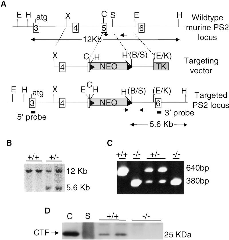
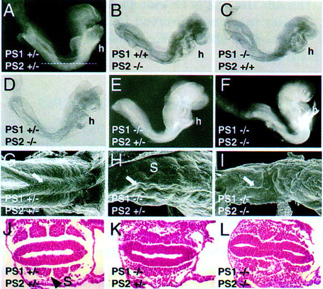
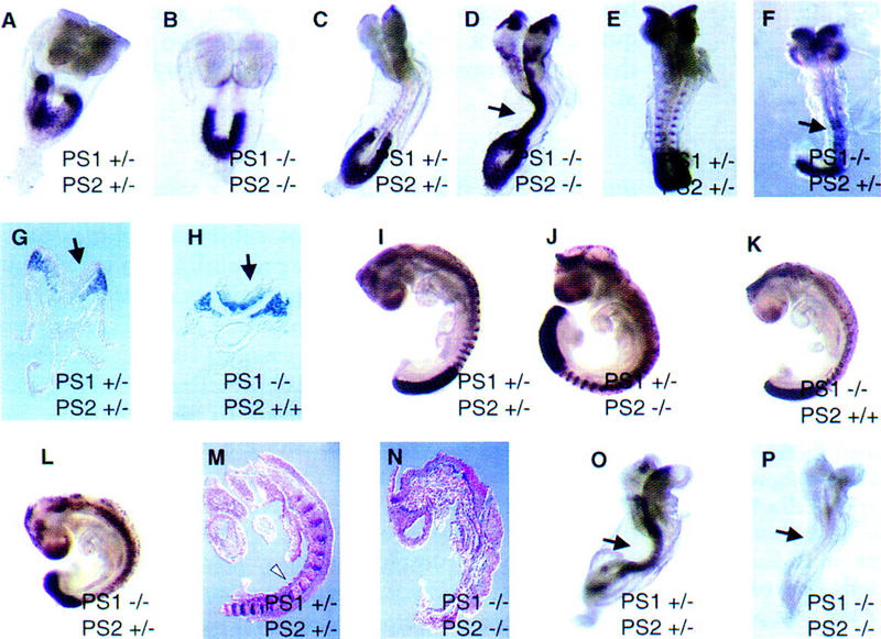
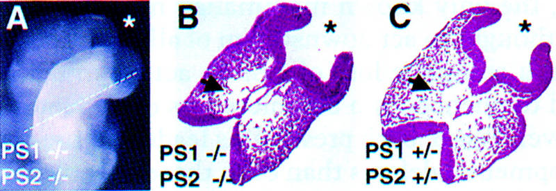
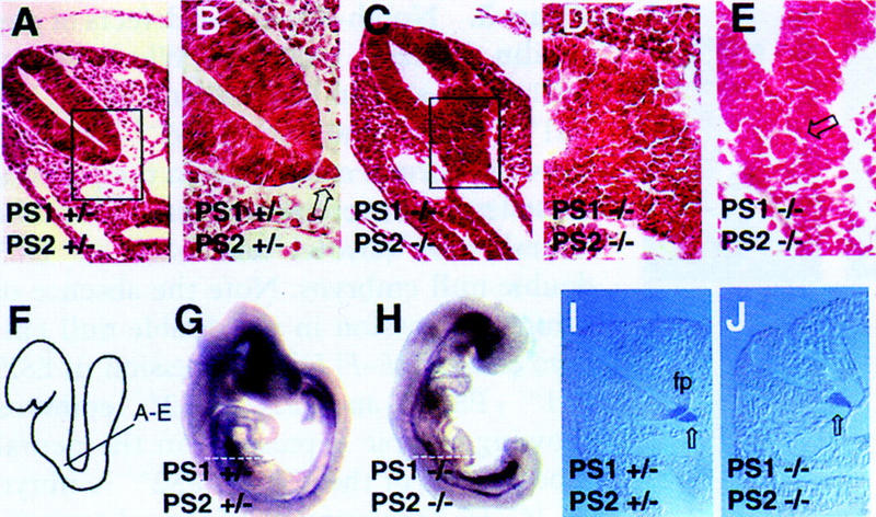
Genetic studies in worms, flies, and humans have implicated the presenilins in the regulation of the Notch signaling pathway and in the pathogenesis of Alzheimer's Disease. There are two highly homologous presenilin genes in mammals, presenilin 1 (PS1) and presenilin 2 (PS2). In mice, inactivation of PS1 leads to developmental defects that culminate in a perinatal lethality. To test the possibility that the late lethality of PS1-null mice reflects genetic redundancy of the presenilins, we have generated PS2-null mice by gene targeting, and subsequently, PS1/PS2 double-null mice. Mice homozygous for a targeted null mutation in PS2 exhibit no obvious defects; however, loss of PS2 on a PS1-null background leads to embryonic lethality at embryonic day 9.5. Embryos lacking both presenilins, and surprisingly, those carrying only a single copy of PS2 on a PS1-null background, exhibit multiple early patterning defects, including lack of somite segmentation, disorganization of the trunk ventral neural tube, midbrain mesenchyme cell loss, anterior neuropore closure delays, and abnormal heart and second branchial arch development. In addition, Delta like-1 (Dll1) and Hes-5, two genes that lie downstream in the Notch pathway, were misexpressed in presenilin double-null embryos: Hes-5 expression was undetectable in these mice, whereas Dll1 was expressed ectopically in the neural tube and brain of double-null embryos. We conclude that the presenilins play a widespread role in embryogenesis, that there is a functional redundancy between PS1 and PS2, and that both vertebrate presenilins, like their invertebrate homologs, are essential for Notch signaling.


Figures

Figure 1
Figure 1
Disruption of the murine PS2 locus by homologous recombination in ES cells. (A) Map of the murine PS2 locus showing the major start of translation in exon 3. Southern analysis using genomic DNA digested with HindIII followed by hybridization with 3′ external or 5′ external probes (█), would yield a 12-kb fragment from the untargeted locus and a 5.6-kb from the targeted locus. Primers (arrows) designed to amplify both the unmodified and the modified loci were used for genotyping by PCR. (NEO) PGK–Neomycin resistance gene; loxP sites (▸) flank the NEO gene; (TK) thymidine kinase gene; (E) EcoRI, (H) HindIII, (X) XbaI, (C) ClaI, (S) StuI, (B) BamHI, (K) KpnI. (B) Southern analysis of HindIII-digested genomic DNA from targeted ES cell clones probed with a PS2 3′ external probe, showing the expected sized fragment for the correct integration of the PS2 targeting vector into the murine PS2 locus in two independent cell lines designated by +/−. (C) PCR analysis on tail DNA from weaning-age mice from PS2 +/− intercrosses using primers indicated in A. Note the absence of the PCR fragment corresponding to the wild-type locus in −/− lanes and the strong band from the doubly targeted PS2 loci. Band sizes are indicated at right. (D) Western blot analysis of brain extracts from PS2+/+ and PS2−/− mice blotted with a PS2 antibody. (CTF) Carboxy-terminal fragment); (C) control extract from human HEK 293t cells transfected with an expression construct driving a mutated (VG) PS2 cDNA. (S) standard lane (NEB). The size of the standard band included in the panel is indicated at right.
Figure 2
Figure 2
Phenotype of presenilin-mutant embryos. E8.5 PS1+/−; PS2+/−(A), PS1+/+; PS2−/−, (B) PS1−/−; PS2+/+(C), and PS1+/−; PS2−/− (D) embryos, which look normal. Note the normal development of the heart (h). The line in A shows the approximate position of the sections shown in J–L. E8.5 PS1−/−; PS2+/− (E) and PS1−/−; PS2−/− (F) embryos, which show no somite segmentation (see K,L), an underdeveloped heart (h, see Fig. 5) and an open anterior neuropore (see Fig. 4). (G–I) SEM of E9 PS1+/−; PS2+/− (G), PS1−/−; PS2+/−(H), and PS1−/−; PS2−/− (I) embryos, showing the neural tube (arrow). Tail bud is on the left. (S) Somites. Transverse sections through E8.5 PS1+/−; PS2+/− (J), PS1−/−; PS2+/−, and PS1−/−; PS2−/− embryos, showing disorganization of the trunk paraxial mesoderm and a kinked neural tube in the presenilin mutants, whereas segmented somites (S, arrowhead) are evident in the PS1+/−; PS2+/− embryo (J).
Figure 3
Figure 3
Notch-associated defects of presenilin mutant embryos. Whole-mount Dll1 expression in E8 (A,B) and E8.5 (C,D) PS1+/−; PS2+/−, and PS1−/−; PS2−/− embryos showing normal expression in the paraxial mesoderm, but ectopic upregulation in the neural tube (arrow) and head of E8.5 double-null embryos. Note the absence of somite expression in the double-null embryo at E8.5. (E–F) Dll1 expression in E8.5 PS1+/−; PS2+/−, and PS1−/−; PS2+/− embryos showing ectopic expression in the neural tube (arrow) of the PS1−/−; PS2+/− embryo (F). (G–H) Transverse sections taken from the tail region of E8.5 double heterozygous and PS1-null embryos hybridized with Dll1 showing expression in the paraxial mesoderm and the neural tube (arrow) of the PS1-null mutant. (I–L) Dll1 expression in E9 presenilin mutant embryos showing PS1−/−; PS2+/− and PS1-null embryos that did not show ectopic expression in the neural tube (K,L). Note the irregularity of Dll1 signal in the presumptive segmentalplate of the PS1−/−; PS2+/− embryo (L).(M–N) UncX4.1 expression in presenilin-mutant embryos. E9 embryos were purposely overstained to ensure detection of any signal on sections from double-null embryos. Saggital sections show UncX4.1 expression in the posterior somitic compartments (white arrowhead) of the normal embryo (M) and no expression in the presenilin double-null embryo (N). (O) Hes-5 expression in the neural tube (arrow) of a normal E8.5 embryo. (P) Absence of Hes-5 expression in an E8.5 presenilin double-null embryo (neural tube is denoted with arrow).
Figure 4
Figure 4
Brain abnormalities in presenilin double-null embryos. (A) Close-up of the head region of an E8.5 double-null embryo showing an open anterior neuropore and an apparent expanded forebrain (*). The line demarcates the approximate position of the section shown in B. (B) Section through the head of a PS1−/−; PS2−/− embryo, which shows loss of mesenchyme cells (arrow). (*) Forebrain. (C) Corresponding section taken from a normal embryo.
Figure 5
Figure 5
Heart and second branchial arch defects of presenilin-mutant embryos. (A) SEM of an E9 PS1+/−; PS2−/− embryo that looks normal. Note the looped heart (solid arrow), normal second branchial arch (open arrow), and closed anterior neural folds (arrowhead). (B,C) S.E.M of E9 PS1−/−; PS2+/− (B) and PS1−/−; PS2−/− (C) embryos exhibiting small, unlooped hearts (solid arrows), absence of second branchial arch development (open arrows), and open neural folds (arrowheads). SEM were photographed at 130× magnification. (D–F) Transverse sections through E8.5 presenilin mutants showing small unlooped hearts (arrows) in the double-null (F) and PS1−/−; PS2+/− (E) embryos.
Figure 6
Figure 6
Neural tube defects of presenilin mutant embryos. (A) Transverse section through a neural tube of a PS1+/−; PS2+/− embryo. The box demarcates the area magnified in the next panel. (B) The boxed region of the dorsal neural tube shown in A at 100× showing ordered columnar morphology of cells. Note the notochord (open arrow) in proximity of the floor plate. (C) The ventral neural tube of a PS1−/−; PS2−/− embryo is disorganized. No notochord is visible, but other sections showed notochord (see E). The boxed region is magnified in the next panel. D Section shown in C is shown at 100×. (E) Transverse section through a PS1−/−; PS2−/− embryo, showing the presence of a notochord (open black arrow), which is surrounded by disorganized cells. (F) Diagram of the approximate position from which the sections shown in (A–E) were derived. E9 PS1+/−; PS2+/− (G) and PS1−/−; PS2−/− (H) embryos hybridized with a probe to Shh. Note the strong expression of Shh in the brain of the double-null embryo (H). The broken lines indicate the position of the sections shown in I–J. (I) Shh expression in the notochord (open arrow) and floor plate (fp) in the trunk region of a normal embryo. (J) Shh expression is detected only in the notochord, and not in the floor plate, in the trunk region of a presenilin double-null embryo.

Similar articles

Cited by

References

    1. Akanawa C, Sasai Y, Nakanishi S, Kageyama R. Molecular characterization of a rat negative regulator with a basic helix-loop-helix structure predominantly expressed in the developing nervous system. J Biol Chem. 1992;267:21879–21885. - PubMed
    1. Anafi M, Kiefer F, Gish GD, Mbamalu G, Iscove NN, Pawson T. SH2/SH3 adaptor proteins can link tyrosine kinases to a ste20-related protein kinase, HPK1. J Biol Chem. 1997;272:27804–27811. - PubMed
    1. Artavanis-Tsakonas S, Rand MD, Lake RJ. Notch signaling: Cell fate control and signal integration in development. Science. 1999;284:770–776. - PubMed
    1. Baumeister R, Leimer U, Zweckbronner I, Jakubek C, Grunberg J, Haass C. Human presenilin-1, but not familial Alzheimer's disease (FAD) mutants, facilitate Caenorhabditis elegans Notch signalling independently of proteolytic processing. Genes Funct. 1997;1:149–159. - PubMed
    1. Berezovska O, Xia MQ, Hyman BT. Notch is expressed in adult brain, is coexpressed with presenilin-1, and is altered in Alzheimer disease. J Neuropathol Exp Neurol. 1998;57:738–745. - PubMed

Publication types