. 1999 Nov 1;13(21):2889-903.
doi: 10.1101/gad.13.21.2889.

Double-strand-break repair recombination in Escherichia coli: physical evidence for a DNA replication mechanism in vivo

Affiliations

Double-strand-break repair recombination in Escherichia coli: physical evidence for a DNA replication mechanism in vivo

M R Motamedi et al. Genes Dev. .

Abstract

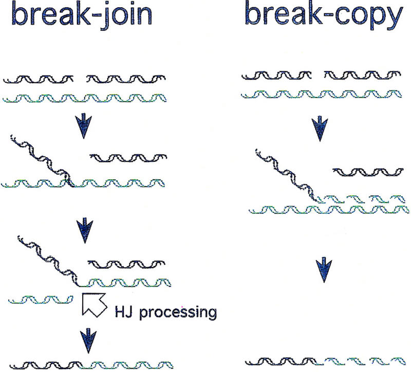
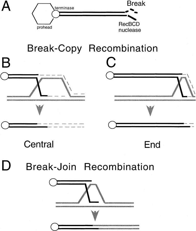
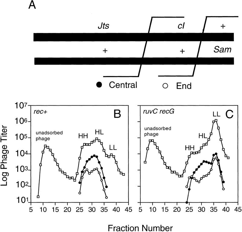
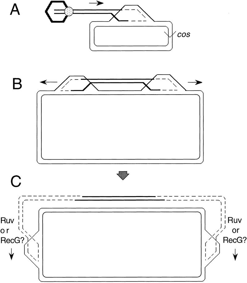
DNA double-strand-break repair (DSBR) is, in many organisms, accomplished by homologous recombination. In Escherichia coli DSBR was thought to result from breakage and reunion of parental DNA molecules, assisted by known endonucleases, the Holliday junction resolvases. Under special circumstances, for example, SOS induction, recombination forks were proposed to initiate replication. We provide physical evidence that this is a major alternative mechanism in which replication copies information from one chromosome to another generating recombinant chromosomes in normal cells in vivo. This alternative mechanism can occur independently of known Holliday junction cleaving proteins, requires DNA polymerase III, and produces recombined DNA molecules that carry newly replicated DNA. The replicational mechanism underlies about half the recombination of linear DNA in E. coli; the other half occurs by breakage and reunion, which we show requires resolvases, and is replication-independent. The data also indicate that accumulation of recombination intermediates promotes replication dramatically.


Figures

Figure 1
Figure 1
Two early general models for homologous recombination (adapted from Meselson and Weigle 1961). Dashed helices represent newly synthesized DNA. Solid helices represent “old” parental DNA. HJ processing (open arrow) indicates action of HJ resolution proteins, including an endonucleolytic cleavage (such as RuvC performs) to break the invaded (green) molecule and allow its ligation to the black fragment. No strand polarities are shown because specific polarities are not implied by either model (see Discussion).
Figure 2
Figure 2
Design of λ crosses used to measure the frequency of recombination in rec+ and ruv recG cells. (A) The strategy for this assay is described in the text (and Razavy et al. 1996). This general diagram shows all of the relevant genetic markers used. The open box (left) represents either of two different deletions (Δb527 or Δb2; Materials and Methods) both starting from the core att site and removing DNA to its left. The solid box represents a deletion/substitution (bio1) starting from the core att site and removing DNA to its right, resulting in a net loss of ∼2 kb of DNA. The arrow indicates the direction of the Chi sequence; + indicates the wild-type copy of the S gene; the other parent carries Sam7. Crosses performed (Fig. 3) varied the presence/absence of the Chi site, Chi+C, and of the nin deletion, Δnin5. All phage are red gam, the top phages by carrying red3 gam210 mutant alleles and the bottom phages by virtue of the bio1 substitution. (B) A representative cesium formate equilibrium density gradient of a cross progeny showing the denser peak formed by site-specific recombination, which contains neither Δ nor bio1 net deletions (fractions 15–20). The next lighter peak (fractions 21–25) includes the top parental phage (A) plus its S+ recombinant derivatives. (□,●) “Total phage” (Sam7 and S+) and S+ recombinants, respectively. These plaques were assayed on SuIII+ recA (for total phage) and SuII+ recA (for S+ recombinants) cells, which do not allow plaque formation of phage with the bio substitution but do allow the gam (amber)210 carriers to form plaques (Materials and Methods). Thus, we do not see the double-deletion (Δbio1) site-specific recombinant peak. To calculate the frequency of λS+ homologous recombinants among site-specific recombinants, the titer of λS+ in each fraction (15–20) is divided by the total titer in that fraction, and the mean ± s.d. for all the fractions in the peak is expressed as a percentage (Fig. 3). The data shown are from a cross in rec+ SMR632 cells using Chio nin+ phage (Materials and Methods).
Figure 3
Figure 3
RecBCD-pathway recombination of λ in the absence of RuvC and RecG. The three graphs summarize the results of three different experimental designs, measuring the efficiency of λ recombination in rec+ and ruvC recG cells. For each design, we used a different set of phages: nin+ (left), Δnin (middle), and Δnin Chi+/o (right). Each bar represents the mean percentage of homologous recombination among site-specific Int-mediated recombinants (±s.d.; calculated as described in Fig. 2). Three independent experiments were performed for nin+ and Δnin crosses. Two experiments were performed for the Δnin Chi+/o cross. The deletion Δnin shortens the DNA segment whose recombination is assayed (see Fig. 2) and therefore necessarily decreases the percent recombination relative to nin+ crosses. Thus, the important comparison for both nin+ and Δnin crosses is between presence or absence of RuvC RecG in each.
Figure 4
Figure 4
λ progeny formation in the absence of DNA replication requires RuvC/RecG. λ progeny formation was used to assay recombination. Replication was blocked by infecting cells that carry a temperature sensitive allele of the dnaE gene with density-labeled λ (λSR27) at 43.5°C, at which temperature we obtain a complete replication block. These graphs show the titers of λ in the fractions of a density gradient obtained following each infection. The densest fractions are to the left on each graph. The first peak in all gradients con-tains unadsorbed λ. These phage carry heavy protein coats and HH DNA. They did not enter the light cells and, therefore, are not part of the λ progeny. These serve as a density reference marker. (A) Density gradient of infection in rec+ cells. Two peaks are apparent. The second peak contains λ progeny that have entered the cell, recombined, and packaged. These carry light protein coats and HH DNA. No other peaks are detected because of the replication block. (B) ruvC recG cells. Few or no λ progeny are detected. (C) recA cells. Few or no λ progeny are detected relative to the unadsorbed phage peak.
Figure 5
Figure 5
Predictions of break–copy and break–join recombination models. The thick lines represent parental DNA (black and gray). The thin broken lines represent newly synthesized DNA. Specific strand polarities are not indicated because no specificities are implied by either model depicted (see Discussion). (A) The phage λ DNA molecule is linearized during DNA packaging by the endonuclease terminase (○), which remains bound to the λ left end after DNA cleavage (Kobayashi et al. 1982, 1983). [Hexagon represents the phage prohead attached to terminase during packaging and concurrent DSBR recombination (Kobayashi et al. 1984).] Only the right end is available for DSBR (Kobayashi et al. 1982, 1983), which begins with degradation leftward by RecBCD exonuclease (for review, see Kowalczykowski et al. 1994; Myers and Stahl 1994). [Note that Chi sites (not shown) are recombination hot spots in this pathway because when RecBCD reaches Chi, Chi decreases RecBCD nuclease activity allowing the DNA there to recombine (for review, see Myers and Stahl 1994; Rosenberg and Motamedi 1999)]. In a break–copy process (B,C), the degraded right end initiates a replication fork. Semiconservative replication of density-labeled DNA to the end of the chromosome followed by the conservative segregation of the new strands (shown) would produce recombinant molecules with the following densities: (C) End recombinants inherit mostly parental DNA and would be expected to band in or near the HH peak in a density transfer experiment (see Fig. 6B,C). (B) Central recombinants would contain roughly half parental and half newly synthesized DNA and would band in the HL peak in a density transfer experiment (see Fig. 6B,C). (D) In break–join, recombination intermediates are resolved by the HJ processing systems. The recombinant molecules inherit only atoms from parental DNA; no new synthesis is required to complete the recombination reaction. These central recombinants would fall into the first few fractions of the HH peak in a density transfer experiment (see Fig. 6B).
Figure 6
Figure 6
Extent of DNA replication in central and right-end λ recombinants in crosses with some replication allowed in rec+ and ruv recG cells. These crosses were conducted under partial replication block (Materials and Methods) to allow visualization of any break–copy recombinants. [If full replication block is used, no HH peak is visible for ruv recG (Fig. 4B).] (A) The relevant genotypes of phages used in this experiment. These phage (Sawitzke and Stahl 1997, Materials and Methods) carry the nin5 deletion and are marked to allow selection of J+ S+ recombinants from which central (J+ cI S+, clear, ●) and right end (J+ cI+ S+, turbid, ○) recombinants are enumerated. (B,C) Density-labeled phages were allowed to recombine under partial replication block and the progenies centrifuged to equilibrium in cesium formate density gradients, which were fractionated. Note that the progenies band into unreplicated, HH, and replicated, HL and LL peaks. Total λ (□) and J+ S+ recombinants were assayed (Materials and Methods), and central (●) and right end (○) recombinants were counted. The first peak (leftward) in these experiments represents unadsorbed phage (heavy coats and HH DNA), which are not part of the λ progeny. (B) Density gradient of the λ cross in rec+ cells. (C) Density gradient of the λ cross in ruvC recG cells.
Figure 7
Figure 7
Break–copy models illustrated for RecBCD-mediated recombination of λ (A) and E. coli (B,C) genomes. The hexagon represents the prohead during λ DNA packaging from cos to cos. The ball represents the terminase protein that linearizes λ, binds the prohead, and packages the DNA.

References

    1. Anderson DG, Kowalczykowski SC. The recombination hot spot chi is a regulatory element that switches the polarity of DNA degradation by the RecBCD enzyme. Genes & Dev. 1997;11:571–581. - PubMed
    1. Appleyard RK. Segregation of new lysogenic types during growth of a doubly lysogenic strain derived from Escherichia coli K12. Genetics. 1954;39:440–452. - PMC - PubMed
    1. Bosco G, Haber JE. Chromosome break-induced DNA replication leads to nonreciprocal translocations and telomere capture. Genetics. 1998;150:1037–1047. - PMC - PubMed
    1. Bull H, Hayes S. The grpD55 locus of Escherichia coli appears to be an allele of dnaB. Mol Gen Genet. 1996;252:755–760. - PubMed
    1. Cairns J, Foster PL. Adaptive reversion of a frameshift mutation in Escherichia coli. Genetics. 1991;128:695–701. - PMC - PubMed

Publication types

LinkOut - more resources