. 1999 Nov;121(3):715-22.
doi: 10.1104/pp.121.3.715.

Expression of endoxyloglucan transferase genes in acaulis mutants of Arabidopsis

Affiliations

Expression of endoxyloglucan transferase genes in acaulis mutants of Arabidopsis

T Akamatsu et al. Plant Physiol. 1999 Nov.

Abstract

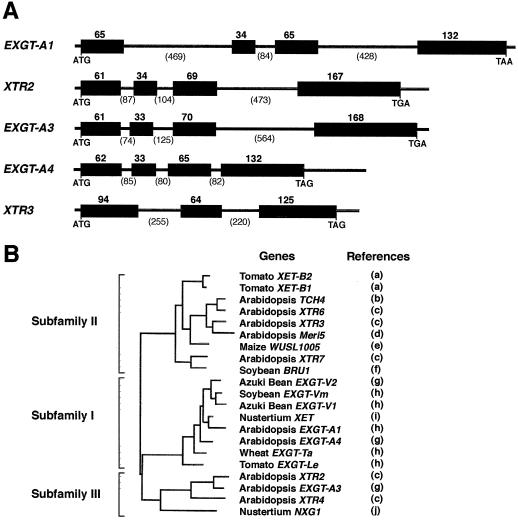
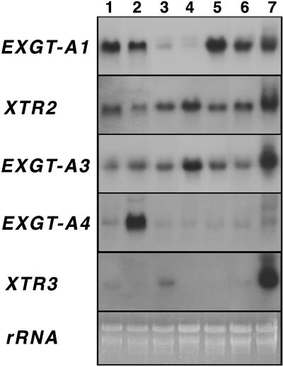
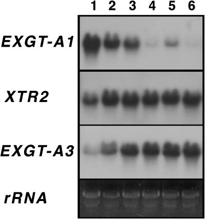
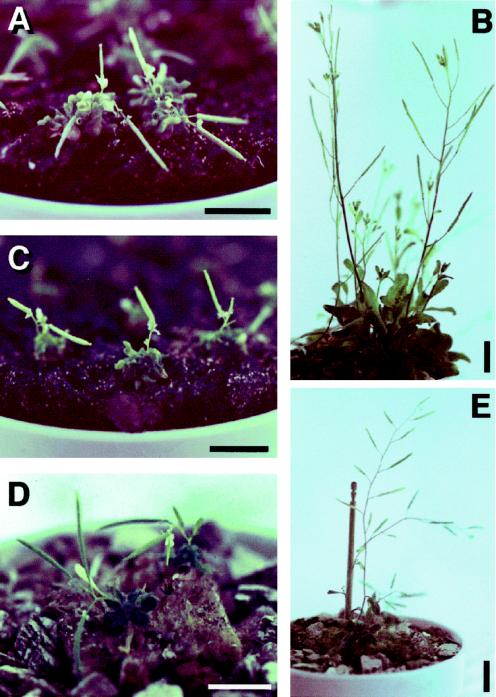
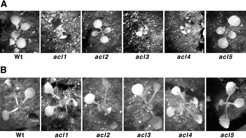
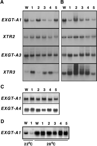
A mutant of Arabidopsis with reduced internodal cell length, acaulis5 (acl5), has recently been shown to have reduced transcript levels of a gene for endoxyloglucan transferase, EXGT-A1 (Y. Hanzawa, T. Takahashi, Y. Komeda [1997] Plant J 12: 863-874). In the present study, we cloned genomic fragments of five members of the EXGT gene family, EXGT-A1, EXGT-A3, EXGT-A4, XTR2, and XTR3, and examined their expression in the wild type and in a series of acl mutants. In wild-type plants, the EXGT-A3 gene showed higher expression in lower internodes (internodes between nodes bearing axillary shoots) than in upper and young internodes, in which EXGT-A1 was highly expressed. EXGT-A4 was preferentially expressed in roots and XTR3 in siliques. The XTR2 gene was constitutively expressed. In acl1, acl3, and acl4 mutants, which have a severe defect in leaf expansion as well as in internode elongation, the EXGT-A1 gene showed reduced levels of expression before bolting of plants. In contrast, XTR3 was increased in these mutant seedlings. Reduction of EXGT-A1 expression was also detected after bolting of all acl mutants except acl2, whose growth defect is restricted to lower internodes. These results suggest the involvement of each EXGT in different aspects of organ development.


Figures

Figure 1
Figure 1
Comparison of EXGT genes. A, Genomic structure of the EXGT genes cloned in this study. Protein coding regions are shown by black boxes with the number of amino acid residues encoded by each exon. Numbers in parentheses indicate the number of nucleotides for intron. Intron splice sites in genomic sequences were deduced by comparison with their corresponding cDNA sequences, EXGT-A1 (Okazawa et al., 1993; accession no. D16454), XTR2 (Xu et al., 1996; accession no. U43487), EXGT-A3 (Nishitani, 1997; accession no. D63509), EXGT-A4 (Nishitani, 1997; accession no. AB026486), and XTR3 (Xu et al., 1996; accession no. U43485). The accession numbers for genomic sequences determined in this study are AF163819 (EXGT-A1), AF163820 (XTR2), AF163821 (EXGT-A3), AF163822 (EXGT-A4), and AF163823 (XTR3), respectively. B, Phylogenetic relationship between the Arabidopsis and other EXGT-related protein sequences. The entire deduced amino acid sequences were compared using the malign program of DNA Data Bank of Japan (Nishitani, 1997). References: a, Arrowsmith and de Silva (1995); b, Xu et al. (1995); c, Xu et al. (1996); d, Medford et al. (1991); e, Saab and Sachs (1995); f, Zurek and Clouse (1994); g, Nishitani (1997); h, Okazawa et al. (1993); i, Rose et al. (1996); and j, de Silva et al. (1993).
Figure 2
Figure 2
Analysis of the expression of EXGT genes in different organs. Total RNA (10 μg per lane) was prepared from 7-d-old seedlings (lane 1), roots (lane 2), rosette leaves (lane 3), internodes between nodes bearing axillary shoots (lane 4), internodes between nodes bearing flowers (lane 5), flower buds (lane 6), and siliques (lane 7).
Figure 3
Figure 3
Analysis of the expression of EXGT genes during internode elongation. Total RNA (10 μg per lane) was prepared from internodes between nodes bearing flowers (lanes 1, 3, and 5) and internodes between nodes bearing axillary shoots (lanes 2, 4, and 6). Tissues were harvested at 5 d (lanes 1 and 2), 10 d (lanes 3 and 4), and 15 d (lanes 5 and 6) after bolting.
Figure 4
Figure 4
Morphology of adult flowering plants with acl mutations. Plants were grown at 22°C under continuous light for 40 d. A, acl1-2; B, acl2-1; C, acl3-1; D, acl4-1; E, acl5-1. Scale bars = 1 cm.
Figure 5
Figure 5
Morphology of 10-d-old wild-type and acl seedlings. Plants were grown under continuous light at 22°C (A) or 28°C (B).
Figure 6
Figure 6
Analysis of the expression of EXGT genes in acl mutants. Total RNA (10 μg per lane) was prepared from aerial tissues of 7-d-old seedlings (A and D) and 30-d-old flowering plants (B) and from root tissues of 7-d-old seedlings (C). Plants were grown at 22°C (A–C) or at the indicated temperature (D). Lanes W, Wild type; lanes 1, acl1-2; lanes 2, acl2-1; lanes 3, acl3-1; lanes 4, acl4-1; lanes 5, acl5-1.

References

    1. Antosiewicz DM, Purugganan MM, Polisensky DH, Braam J. Cellular localization of Arabidopsis xyloglucan endotransglycosylase-related proteins during development and after wind stimulation. Plant Physiol. 1997;115:1319–1328. - PMC - PubMed
    1. Arrowsmith DA, de Silva J. Characterisation of two tomato fruit-expressed cDNAs encoding xyloglucan endo-transglycosylase. Plant Mol Biol. 1995;28:391–403. - PubMed
    1. Bell CJ, Ecker JR. Assignment of 30 microsatellite loci to the linkage map of Arabidopsis. Genomics. 1994;19:137–144. - PubMed
    1. Carpita NC, Gibeaut DM. Structural models of primary cell walls in flowering plants: consistency of molecular structure with the physical properties of the walls during growth. Plant J. 1993;3:1–30. - PubMed
    1. Cosgrove DJ. Cell wall loosening by expansins. Plant Physiol. 1998;118:333–339. - PMC - PubMed

Publication types