

Peroxisomal NADP-Dependent Isocitrate Dehydrogenase. Characterization and Activity Regulation during Natural Senescence
- PMID: 10557241
- PMCID: PMC59455
- DOI: 10.1104/pp.121.3.921
Peroxisomal NADP-Dependent Isocitrate Dehydrogenase. Characterization and Activity Regulation during Natural Senescence
Abstract
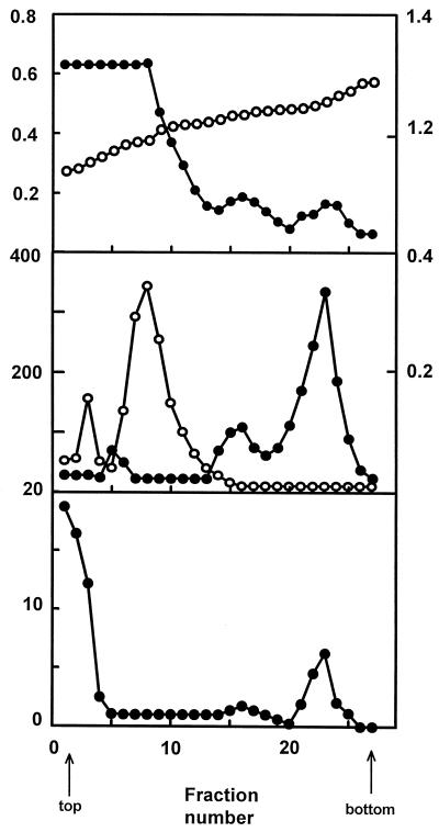
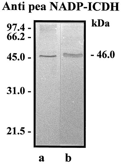
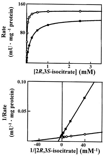
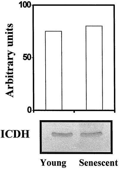
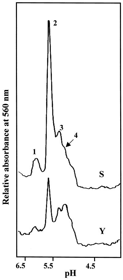
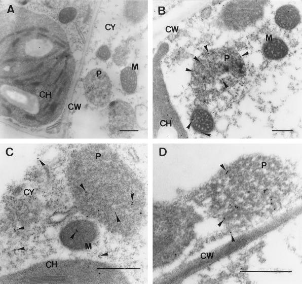
The peroxisomal localization and characterization of NADP-dependent isocitrate dehydrogenase (perICDH) in young and senescent pea (Pisum sativum) leaves was studied by subcellular fractionation, kinetic analysis, immunoblotting, and immunoelectron microscopy. The subunit molecular mass for perICDH determined by immunoblotting was 46 kD. By isoelectric focusing (IEF) of the peroxisomal matrix fraction, the NADP-ICDH activity was resolved into four isoforms, perICDH-1 to perICDH-4, with isoelectric points (pIs) of 6.0, 5.6, 5.4, and 5.2, respectively. The kinetic properties of the NADP-ICDH in peroxisomes from young and senescent pea leaves were analyzed. The maximum initial velocity was the same in peroxisomes from young and senescent leaves, while the Michaelis constant value in senescent leaf peroxisomes was 11-fold lower than in young leaf peroxisomes. The protein levels of NADP-ICDH in peroxisomes were not altered during senescence. The kinetic behavior of this enzyme suggests a possible fine control of enzymatic activity by modulation of its Michaelis constant during the natural senescence of pea leaves. After embedding, electron microscopy immunogold labeling of NADP-ICDH confirmed that this enzyme was localized in the peroxisomal matrix. Peroxisomal NADP-ICDH represents an alternative dehydrogenase in these cell organelles and may be the main system for the reduction of NADP to NADPH for its re-utilization in the peroxisomal metabolism.
Figures
Similar articles
-
Peroxisomal NADP-isocitrate dehydrogenase is required for Arabidopsis stomatal movement.Protoplasma. 2016 Mar;253(2):403-15. doi: 10.1007/s00709-015-0819-0. Epub 2015 Apr 19. Protoplasma. 2016. PMID: 25894616
-
A dehydrogenase-mediated recycling system of NADPH in plant peroxisomes.Biochem J. 1998 Mar 1;330 ( Pt 2)(Pt 2):777-84. doi: 10.1042/bj3300777. Biochem J. 1998. PMID: 9480890 Free PMC article.
-
Activity and gene expression analysis of the NADP-dependent isocitrate dehydrogenase (NADP-ICDH) through pepper fruit ripening and its modulation by nitric oxide (NO). Molecular characterization of the peroxisomal isozyme.Plant Sci. 2024 Dec;349:112269. doi: 10.1016/j.plantsci.2024.112269. Epub 2024 Sep 21. Plant Sci. 2024. PMID: 39313003
-
Peroxisomal plant metabolism - an update on nitric oxide, Ca2+ and the NADPH recycling network.J Cell Sci. 2018 Jan 29;131(2):jcs202978. doi: 10.1242/jcs.202978. J Cell Sci. 2018. PMID: 28775155 Review.
-
Control of isocitrate dehydrogenase catalytic activity by protein phosphorylation in Escherichia coli.J Mol Microbiol Biotechnol. 2005;9(3-4):132-46. doi: 10.1159/000089642. J Mol Microbiol Biotechnol. 2005. PMID: 16415587 Review.
Cited by
-
Tricarboxylic Acid (TCA) Cycle Intermediates: Regulators of Immune Responses.Life (Basel). 2021 Jan 19;11(1):69. doi: 10.3390/life11010069. Life (Basel). 2021. PMID: 33477822 Free PMC article. Review.
-
Protein tyrosine nitration in pea roots during development and senescence.J Exp Bot. 2013 Feb;64(4):1121-34. doi: 10.1093/jxb/ert006. Epub 2013 Jan 28. J Exp Bot. 2013. PMID: 23362300 Free PMC article.
-
Peroxisomal NADP-isocitrate dehydrogenase is required for Arabidopsis stomatal movement.Protoplasma. 2016 Mar;253(2):403-15. doi: 10.1007/s00709-015-0819-0. Epub 2015 Apr 19. Protoplasma. 2016. PMID: 25894616
-
Phytochemicals: "A Small Defensive Advantage for Plants and Fungi; a Great Remedy for the Health of Mankind".Molecules. 2021 Oct 12;26(20):6159. doi: 10.3390/molecules26206159. Molecules. 2021. PMID: 34684740 Free PMC article.
-
LncRNA IDH1-AS1 links the functions of c-Myc and HIF1α via IDH1 to regulate the Warburg effect.Proc Natl Acad Sci U S A. 2018 Feb 13;115(7):E1465-E1474. doi: 10.1073/pnas.1711257115. Epub 2018 Jan 29. Proc Natl Acad Sci U S A. 2018. PMID: 29378948 Free PMC article.
References
-
- Aebi H. Catalase in vitro. Methods Enzymol. 1984;105:121–126. - PubMed
-
- Attucci S, Rivoal J, Brouquisse R, Carde JP, Pradet A, Raymond P. Characterization of a mitochondrial NADP-dependent isocitrate dehydrogenase in axes of germinating sunflower seeds. Plant Sci. 1994;102:49–59.
-
- Barroso JB. Influencias nutricionales y de la edad sobre el comportamiento cinético de los sistemas productores de NADPH en diferentes tejidos de la trucha arco-iris (Oncorhynchus mykiss). PhD thesis. Spain: University of Granada; 1993.
-
- Bradford MM. A rapid and sensitive method for the quantitation of microgram quantities of protein utilizing the principle of protein-dye binding. Anal Biochem. 1976;72:248–254. - PubMed
-
- Breidenbach RW, Beevers H. Association of the glyoxylate cycle enzymes in a novel subcellular particle from castor bean endosperm. Biochem Biophys Res Commun. 1967;27:462–469. - PubMed
LinkOut - more resources
Full Text Sources
