

Rational engineering of a miniprotein that reproduces the core of the CD4 site interacting with HIV-1 envelope glycoprotein
- PMID: 10557278
- PMCID: PMC23905
- DOI: 10.1073/pnas.96.23.13091
Rational engineering of a miniprotein that reproduces the core of the CD4 site interacting with HIV-1 envelope glycoprotein
Abstract
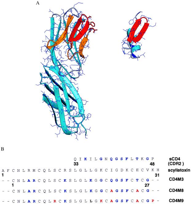
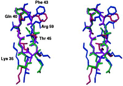
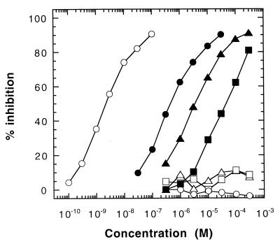
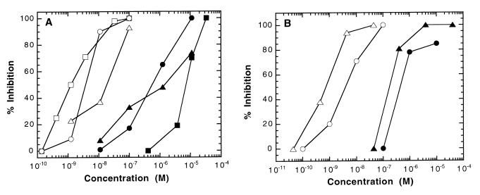
Protein-protein interacting surfaces are usually large and intricate, making the rational design of small mimetics of these interfaces a daunting problem. On the basis of a structural similarity between the CDR2-like loop of CD4 and the beta-hairpin region of a short scorpion toxin, scyllatoxin, we transferred the side chains of nine residues of CD4, central in the binding to HIV-1 envelope glycoprotein (gp120), to a structurally homologous region of the scorpion toxin scaffold. In competition experiments, the resulting 27-amino acid miniprotein inhibited binding of CD4 to gp120 with a 40 microM IC(50). Structural analysis by NMR showed that both the backbone of the chimeric beta-hairpin and the introduced side chains adopted conformations similar to those of the parent CD4. Systematic single mutations suggested that most CD4 residues from the CDR2-like loop were reproduced in the miniprotein, including the critical Phe-43. The structural and functional analysis performed suggested five additional mutations that, once incorporated in the miniprotein, increased its affinity for gp120 by 100-fold to an IC(50) of 0.1-1.0 microM, depending on viral strains. The resulting mini-CD4 inhibited infection of CD4(+) cells by different virus isolates. Thus, core regions of large protein-protein interfaces can be reproduced in miniprotein scaffolds, offering possibilities for the development of inhibitors of protein-protein interactions that may represent useful tools in biology and in drug discovery.
Figures

References
-
- Dalgleish A G, Beverley A C, Clapham P R, Crawford D H, Greaves M F, Weiss R A. Nature (London) 1984;312:763–767. - PubMed
-
- Klatzmann D, Champagne E, Chamaret S, Gruest J, Guetard D, Hercend T, Gluckman J C, Montagnier L. Nature (London) 1984;312:767–768. - PubMed
-
- Wu L, Gerard N P, Wyatt R, Choe H, Parolin C, Ruffing A, Borsetti A, Cardoso A A, Desjardin E, Newman W, et al. Nature (London) 1996;384:179–183. - PubMed
-
- Trkola A, Dragic T, Arthos J, Binley J M, Olson W C, Allaway G P, Cheng-Mayer C, Robinson J, Maddon P J, Moore J P. Nature (London) 1996;384:184–187. - PubMed
-
- Berger E A. AIDS. 1997;11:S3–S16. - PubMed
Publication types
MeSH terms
Substances
Associated data
- Actions
LinkOut - more resources
Full Text Sources
Other Literature Sources
Research Materials
