. 1999 Nov 9;96(23):13264-9.
doi: 10.1073/pnas.96.23.13264.

cDNA microarrays detect activation of a myogenic transcription program by the PAX3-FKHR fusion oncogene

Affiliations

cDNA microarrays detect activation of a myogenic transcription program by the PAX3-FKHR fusion oncogene

J Khan et al. Proc Natl Acad Sci U S A. .

Abstract

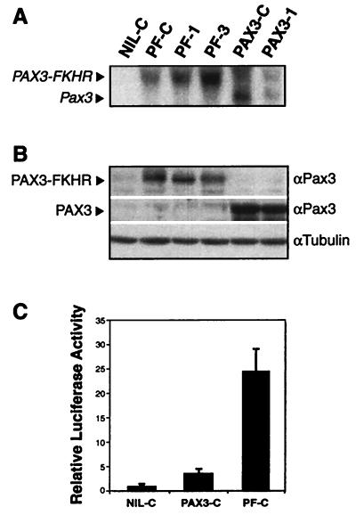
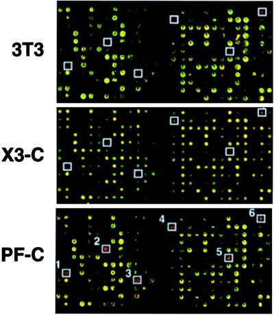
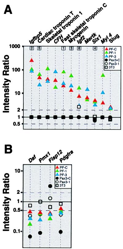
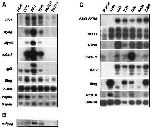
Alveolar rhabdomyosarcoma is an aggressive pediatric cancer of striated muscle characterized in 60% of cases by a t(2;13)(q35;q14). This results in the fusion of PAX3, a developmental transcription factor required for limb myogenesis, with FKHR, a member of the forkhead family of transcription factors. The resultant PAX3-FKHR gene possesses transforming properties; however, the effects of this chimeric oncogene on gene expression are largely unknown. To investigate the actions of these transcription factors, both Pax3 and PAX3-FKHR were introduced into NIH 3T3 cells, and the resultant gene expression changes were analyzed with a murine cDNA microarray containing 2,225 elements. We found that PAX3-FKHR but not PAX3 activated a myogenic transcription program including the induction of transcription factors MyoD, Myogenin, Six1, and Slug as well as a battery of genes involved in several aspects of muscle function. Notable among this group were the growth factor gene Igf2 and its binding protein Igfbp5. Relevance of this model was suggested by verification that three of these genes (IGFBP5, HSIX1, and Slug) were also expressed in alveolar rhabdomyosarcoma cell lines. This study utilizes cDNA microarrays to elucidate the pattern of gene expression induced by an oncogenic transcription factor and demonstrates the profound myogenic properties of PAX3-FKHR in NIH 3T3 cells.


Figures

Figure 1
Figure 1
(A) Northern blot analysis confirms expression of PAX3-FKHR in the lines PF-C, PF-1, and PF-3 and Pax3 in PAX3-C and PAX3–1. (B) Western blot analysis confirms comparable levels of these proteins in accord with the Northern blot data. (C) Relative luciferase activity of PAX3-FKHR (in PF-C) showing ≈6× the transcriptional activity of Pax3 (in PAX3-C), as described (4). The assay was performed according to published protocols (42) using the Pax3 reporter plasmid plucTKCD19.
Figure 2
Figure 2
Representative microarray hybridization of transduced lines. For each experiment, we used the same reference target prepared from cells transduced with empty vector (designated NIL-C). The pseudocolored images represent portions of a microarray with reference target, NIL-C, in green and parental NIH 3T3, PAX3-C, and PF-C in red. Up-regulation of several genes of interest by PAX3-FKHR are boxed (1, Fast skeletal troponin C; 2, Igfbp5; 3, Myogenin; 4, Six1; 5, Cardiac troponin T; 6, Igf2).
Figure 3
Figure 3
(A) Eleven genes induced in PF-C, PF-1, and PF-3. The intensity ratio (log10 scale) is plotted against the 11 genes induced (at the 99% confidence level in each individual experiment) for all five microarray hybridizations. When the intensity values for both red and green channels for a probe/spot were <1,400, then the ratio of gene expression cannot be determined with precision, and the ratio value is plotted as 1. Boxed numbers correspond to genes from Fig. 2. (B) Four genes down-regulated by PAX3-FKHR, of which three (Daf, Pmx1, and Pdgfra) were also repressed by PAX3. A stringent filter, including only those genes with fluorescence ≥5,000 for either red or green channels, was applied for this analysis.
Figure 4
Figure 4
Northern blot analysis confirms array data. (A) Induction of Six1, Myogenin, MyoD, Igfbp5, Igf2, and Slug by PAX3-FKHR transduction is demonstrated, as is the failure to induce these genes by PAX3. Igf2 expression appears by Northern blotting to be suppressed by PAX3. Basal c-Met levels in NIH 3T3 cells transduced by the empty virus is high and remains high in the Pax3 and PAX3-FKHR transduced clones. Pdgfra expression is repressed in the clones PF-1, PF-3, PAX3-C, and PAX3–1. (B) Myogenin protein expression is confirmed in the PAX3-FKHR transduced clones PF-C, PF-1, and PF-3 by Western analysis. (C) Northern blot analysis reveals expression of HSIX1, MYOG, IGFBP5, IGF2, Slug, and MERTK in several of the ARMS cell lines (RH3, RH4, RH5, RH28, and RH30). The first lane is normal muscle. A204 is an embryonal rhabdomyosarcoma that lacks the PAX3-FKHR chimeric oncogene.

Similar articles

Cited by

References

    1. Barr F G. Nat Genet. 1998;19:121–124. - PubMed
    1. Galili N, Davis R J, Fredericks W J, Mukhopadhyay S, Rauscher F J, III, Emanuel B S, Rovera G, Barr F G. Nat Genet. 1993;5:230–235. - PubMed
    1. Shapiro D N, Sublett J E, Li B, Downing J R, Naeve C W. Cancer Res. 1993;53:5108–5112. - PubMed
    1. Fredericks W J, Galili N, Mukhopadhyay S, Rovera G, Bennicelli J, Barr F G, Rauscher F J., III Mol Cell Biol. 1995;15:1522–1535. - PMC - PubMed
    1. Bennicelli J L, Fredericks W J, Wilson R B, Rauscher F J, III, Barr F G. Oncogene. 1995;11:119–130. - PubMed