. 1999 Nov 9;96(23):13300-5.
doi: 10.1073/pnas.96.23.13300.

Accumulation of premutagenic DNA lesions in mice defective in removal of oxidative base damage

Affiliations

Accumulation of premutagenic DNA lesions in mice defective in removal of oxidative base damage

A Klungland et al. Proc Natl Acad Sci U S A. .

Abstract

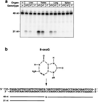
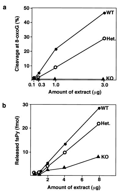
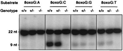
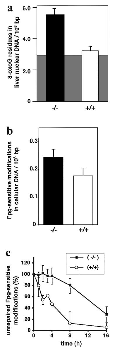
DNA damage generated by oxidant byproducts of cellular metabolism has been proposed as a key factor in cancer and aging. Oxygen free radicals cause predominantly base damage in DNA, and the most frequent mutagenic base lesion is 7,8-dihydro-8-oxoguanine (8-oxoG). This altered base can pair with A as well as C residues, leading to a greatly increased frequency of spontaneous G.C-->T.A transversion mutations in repair-deficient bacterial and yeast cells. Eukaryotic cells use a specific DNA glycosylase, the product of the OGG1 gene, to excise 8-oxoG from DNA. To assess the role of the mammalian enzyme in repair of DNA damage and prevention of carcinogenesis, we have generated homozygous ogg1(-/-) null mice. These animals are viable but accumulate abnormal levels of 8-oxoG in their genomes. Despite this increase in potentially miscoding DNA lesions, OGG1-deficient mice exhibit only a moderately, but significantly, elevated spontaneous mutation rate in nonproliferative tissues, do not develop malignancies, and show no marked pathological changes. Extracts of ogg1 null mouse tissues cannot excise the damaged base, but there is significant slow removal in vivo from proliferating cells. These findings suggest that in the absence of the DNA glycosylase, and in apparent contrast to bacterial and yeast cells, an alternative repair pathway functions to minimize the effects of an increased load of 8-oxoG in the genome and maintain a low endogenous mutation frequency.


Figures

Figure 1
Figure 1
Targeted disruption of the murine OGG1 locus. (a) Physical map of the genomic DNA containing the OGG1 gene. The location of the helix–hairpin–helix motif is indicated; amino acids identical in yeast, human, and murine OGG1 are boxed; the murine amino acid sequence shown is identical to the human sequence except for the two residues underlined; the Lys and Asp residues associated with DNA glycosylase/AP lyase activity are shown with an arrow and a circle, respectively (15, 17). Genomic fragments were subcloned on either side of the Neo gene to generate a construct that deleted ≈4.6 kb including this motif at the targeted locus. Genomic DNA is shown by boxes; vector sequences are shown by a line. Restriction digest with EcoRI gives rise to a ≈12-kb fragment at the wild-type locus and a ≈5-kb fragment at the targeted locus that are detected by hybridization with a 5′ flanking probe (striped box). (b) Representative wild-type (+/+), heterozygous (+/−), and ogg1 null (−/−) live-born F2 progeny genotyped by EcoRI digestion of tailsnip DNA and hybridization with the 5′ probe.
Figure 2
Figure 2
Absence of OGG1 activity in extracts of organs from ogg1 null mice. (a) Nuclear extracts were prepared of the organs indicated from wild-type (+/+), heterozygous (+/−), and ogg1 null (−/−) mice and incubated with a double-stranded oligonucleotide substrate (b) with a 8-oxoG:C mismatch and 32P-labeled at the 5′ end of the 8-oxoG-containing strand. Reaction products were analyzed by PhosphorImager after denaturing PAGE. OGG1 activity excises the 8-oxoG and cleaves the 32P-labeled, 8-oxoG-containing strand (49 nt) 3′ of the lesion; subsequent cleavage of the terminal sugar phosphate by HAP1 endonuclease (36) in the extract produces a 21-nt, 32P-labeled species. Lower molecular weight bands result from exonuclease degradation at the exposed 3′ residues of the 21-nt cleavage product. Levels of two control enzyme activities, uracil-DNA glycosylase and hNth1 (36), were normal in all extracts.
Figure 3
Figure 3
Comparative activity on 8-oxoG and faPy DNA lesions in extracts from ogg1 null mice. Nuclear extracts prepared from testes of wild-type (WT, ●), heterozygous (Het., ○), and ogg1 knockout (KO, ▴) mice were assayed for either cleavage at 8-oxoG:C (a) or release of faPy lesions (b) by using appropriate DNA substrates.
Figure 4
Figure 4
Comparative activity on different 8-oxoG-containing mismatches in extracts from ogg1 null mice. Nuclear extracts prepared from testes of wild-type (+/+), heterozygous (+/−), and ogg1 null (−/−) mice were incubated with double-stranded oligonucleotide substrates containing different residues opposite 8-oxoG (as indicated). The assay was as in Fig. 2, except, in this case, a 22-bp substrate was used and OGG1 activity is indicated by cleavage of the 32P-labeled 8-oxoG-containing strand to produce a 9-nt species.
Figure 5
Figure 5
Apparent steady-state levels and delayed repair of oxidative DNA damage in liver (a) and an MEF cell line (b and c) from ogg1 null mice. (a) Nuclear DNA was isolated from the liver of ogg1−/− null (solid bar) or wild-type OGG1+/+ (open bar) adult mice under conditions designed to minimize artificial background oxidation (shaded area) during the extraction and work-up process (see text), and levels of 8-oxoG were quantitated by HPLC-ECD after enzymatic DNA hydrolysis. (b) Total DNA was isolated from cultured ogg1−/− null (solid bar) or wild-type OGG1+/+ (open bar) MEF cell lines, and levels of Fpg-sensitive modifications were quantitated by alkaline elution. (c) Oxidative DNA damage was induced in the ogg1−/− null (■) or wild-type OGG1+/+ (○) cell line by treatment with a photosensitizer and visible light. The persistence of Fpg-sensitive base modifications was measured (as in b) after various recovery times up to 16 h and corrected for dilution of the induced damage by cell proliferation. Error bars are shown for the SD of the mean from five mice (a) and five (b) or three (c) replica experiments. Note the different scale for y axes in a and b.

References

    1. Ames B N, Shigenaga M K, Hagen T M. Proc Natl Acad Sci USA. 1997;90:7915–7922. - PMC - PubMed
    1. Collins A R. BioEssays. 1999;21:238–246. - PubMed
    1. Grollman A P, Moriya M. Trends Genet. 1993;9:246–249. - PubMed
    1. Ward J F. In: Advances in DNA Damage and Repair. Dizdaroglu M, Karakaya A E, editors. New York: Plenum; 1998. pp. 431–439.
    1. Michaels M L, Miller J H. J Bacteriol. 1992;174:6321–6325. - PMC - PubMed

Publication types