. 1999 Nov 9;96(23):13336-41.
doi: 10.1073/pnas.96.23.13336.

Increased thrombin responsiveness in platelets from mice lacking glycoprotein V

Affiliations

Increased thrombin responsiveness in platelets from mice lacking glycoprotein V

V Ramakrishnan et al. Proc Natl Acad Sci U S A. .

Abstract

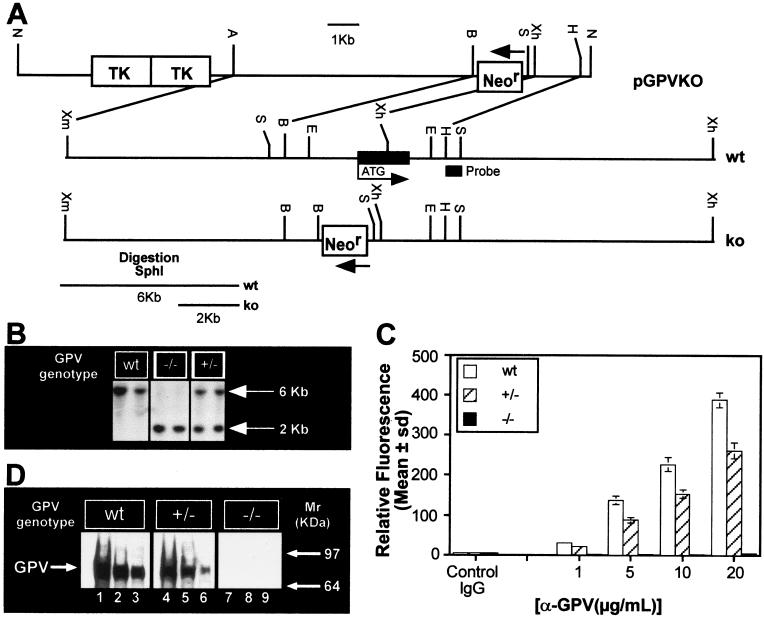
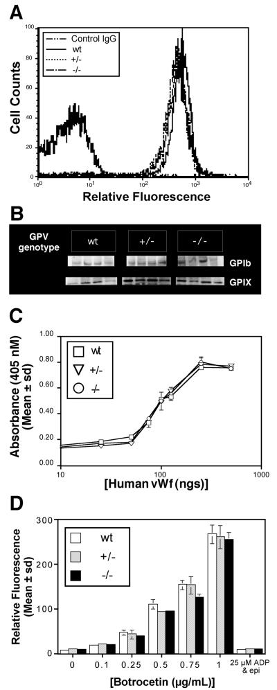
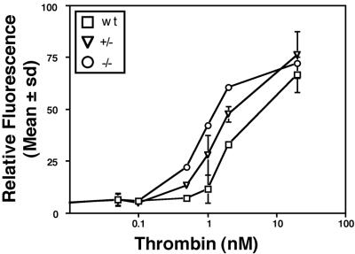
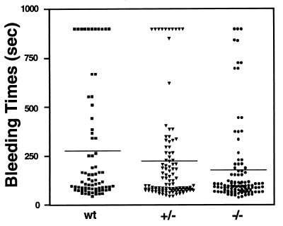
A role for glycoprotein (GP)V in platelet function has been proposed on the basis of observations that GP V is the major thrombin substrate on intact platelets cleaved during thrombin-induced platelet aggregation, and that GP V promotes GP Ib-IX surface expression in heterologous cells. We tested the hypotheses that GP V is involved in thrombin-induced platelet activation, in GP Ib-IX expression, and in other platelet responses by generating GP V null mice. Contrary to expectations, GP V -/- platelets were normal in size and expressed normal amounts of GP Ib-IX that was functional in von Willebrand factor binding, explaining why defects in GP V have not been observed in Bernard-Soulier syndrome, a bleeding disorder caused by a lack of functional GP Ib-IX-V. Moreover, in vitro analysis demonstrated that GP V -/- platelets were hyperresponsive to thrombin, resulting in increased fibrinogen binding and an increased aggregation response. Consistent with these findings, GP V -/- mice had a shorter bleeding time. These data support a role for GP V as a negative modulator of platelet activation. Furthermore, they suggest a new mechanism by which thrombin enhances platelet responsiveness independent of activation of the classical G-protein-coupled thrombin receptors.


Figures

Figure 1
Figure 1
(A) Genomic organization of the mouse GPV gene and generation of the targeting vector. The targeting vector pGPVKO contained a 5′ HR [8 kb; XmaI(blunt)–BamHI fragment of GP V] and a 3′HR (1.4 kb, XhoI–HindIII fragment of GP V) around the Neor cassette, as shown (B = BamHI, E = EcoRI, A = Acc651, H = HindIII, N = NotI, S = SphI, Xh = XhoI, Xm = XmaI). (B) Southern blot analysis of mouse-tail genomic DNA digested with SphI. The probe used is indicated in A. (C) FACS analysis of wt, +/− and −/− platelets. PRP from wt +/− and −/− mice was incubated with the indicated concentrations of Ab #810 and analyzed by flow cytometry. (D) Western analysis of GP V wt, +/− and −/− platelets. WP lysates were electrophoresed, transferred, and incubated with Ab #808 (5 μg/ml) followed by ECL. Lanes 1–3, wt, lanes 4–6, +/−, and lanes 7–9, −/−. Lanes 1, 4, 7: 2 × 10⁷ platelets; lanes 2, 5, 8: 1 × 10⁷ platelets; lanes 3, 6, 9: 5 × 10⁶ platelets. Mouse GP V has a Mr ≈ 89,000 kDa.
Figure 2
Figure 2
(A) Surface expression of GP Ib-IX on GP V wt, +/− and −/− platelets. WP were incubated with Ab #3584 (1:1,000 dilution) and analyzed by flow cytometry. The data are representative of six animals of each genotype. Control IgG is on the left. (B) Western analysis of GP Ib-IX expression on GP V wt, +/− and −/− platelets. Lysed WP (5 × 10⁷) were electrophoresed, transferred, and incubated with Ab#3584 (1:1,000) followed by ECL. Four individual mice of each genotype are shown (C) Binding of GP V wt, +/− and −/− platelets to immobilized human vWf. Pooled WP were incubated as described in Methods. The data shown are the average of duplicates and are representative of three such experiments. (D) Binding of FITC–vWf to GP V wt, +/− and −/− platelets. Pooled PRP was incubated with FITC–vWf and botrocetin and analyzed by flow cytometry. The data are representative of three experiments done in duplicate.
Figure 3
Figure 3
Thrombin-induced FITC–fibrinogen binding in GP V wt, +/− and −/− platelets. WP from individual mice were stimulated with the indicated amounts of thrombin, incubated with FITC-labeled fibrinogen for 30 min, and fixed and analyzed by flow cytometry. The data are representative of three experiments done in duplicate.
Figure 4
Figure 4
(A) Thrombin-induced aggregation in GP V wt +/− and −/− platelets. WP were prepared from six to ten mice of each genotype, which were littermate controls. Platelet aggregation was determined in a lumi-aggregometer. Five experiments were carried out, and four worked in the manner shown. (B) FACS analysis of surface αΙΙbβ3 levels in GP V wt, +/− and −/− platelets. PRP was incubated with Ab #41 (10 μg/ml), and samples were analyzed by flow cytometry. The data are representative of six individual mice per genotype. Control IgG is on the left. (C) Western analysis of total αΙΙbβ3 levels in GP V wt, +/− and −/− platelets. WP lysates were electrophoresed, transferred to nitrocellulose, and incubated with Ab #41 (10 μg/ml) followed by ECL. Four individual mice of each genotype are shown.
Figure 5
Figure 5
Determination of bleeding time. Bleeding time measurements were obtained by using the tail-cut model on littermate mice (wt n = 74; +/− n = 105, −/− n = 106) generated from heterozygous breeding, as described in Methods. Each symbol represents a single animal.

References

    1. Baumgartner H R, Tschopp T B, Weiss H J. Thromb Haemostasis. 1978;39:782–783. - PubMed
    1. Clemetson K J, Clemetson J M. Semin Thromb Hemostasis. 1995;21:130–136. - PubMed
    1. Davies M J. Br Med Bull. 1994;50:789–802. - PubMed
    1. Jang Y, Lincoff A M, Plow E F, Topol E J. J Am Coll Cardiol. 1994;24:1591–1601. - PubMed
    1. Lopez J A, Andrews R K, Afshar-Kharghan V, Berndt M C. Blood. 1998;91:4397–4418. - PubMed

Associated data