. 1999 Nov 9;96(23):13409-14.
doi: 10.1073/pnas.96.23.13409.

Serine racemase: a glial enzyme synthesizing D-serine to regulate glutamate-N-methyl-D-aspartate neurotransmission

Affiliations

Serine racemase: a glial enzyme synthesizing D-serine to regulate glutamate-N-methyl-D-aspartate neurotransmission

H Wolosker et al. Proc Natl Acad Sci U S A. .

Abstract

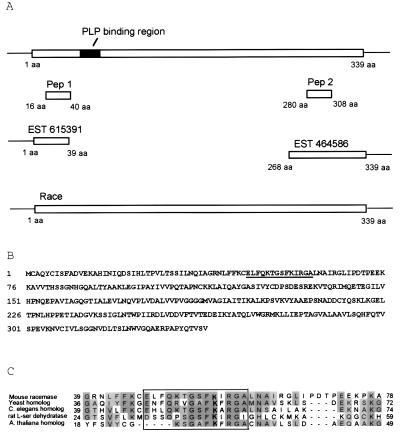
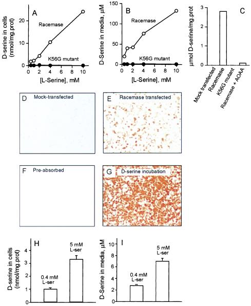
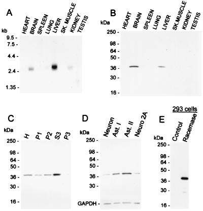
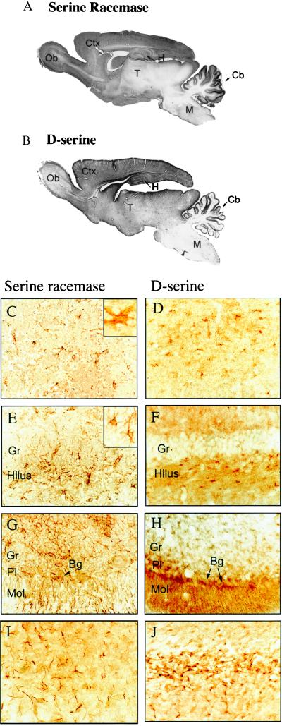
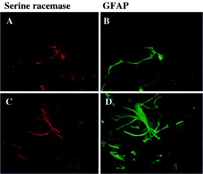
Although D amino acids are prominent in bacteria, they generally are thought not to occur in mammals. Recently, high levels of D-serine have been found in mammalian brain where it activates glutamate/N-methyl-D-aspartate receptors by interacting with the "glycine site" of the receptor. Because amino acid racemases are thought to be restricted to bacteria and insects, the origin of D-serine in mammals has been puzzling. We now report cloning and expression of serine racemase, an enzyme catalyzing the formation of D-serine from L-serine. Serine racemase is a protein representing an additional family of pyridoxal-5' phosphate-dependent enzymes in eukaryotes. The enzyme is enriched in rat brain where it occurs in glial cells that possess high levels of D-serine in vivo. Occurrence of serine racemase in the brain demonstrates the conservation of D-amino acid metabolism in mammals with implications for the regulation of N-methyl-D-aspartate neurotransmission through glia-neuronal interactions.


Figures

Figure 1
Figure 1
Cloning and sequence analysis of mouse serine racemase. (A) Strategy for serine racemase cloning. Two peptides obtained by internal amino acid sequence of purified enzyme (PEP1, HLNIQDSVHLTPVLTSSILNQIAGR; PEP2, LLIEPTAGVGLAAVLSQHFQTVSPEVK) corresponded to mouse ESTs in GenBank. Full-length serine racemase cloned by reverse transcription–PCR from mouse brain has a pyridoxal-5′ phosphate (PLP) binding region. The ORF and stop codon were confirmed by an independent 3′ rapid amplification of cDNA ends (Race) reaction. (B) Amino acid sequence of serine racemase. Underlined sequence corresponds to the amino acid consensus for pyridoxal 5′ phosphate binding (Prosite accession number PS00165). (C) Alignment of proteins exhibiting significant homology to serine racemase in the pyridoxal 5′ phosphate binding region (boxed area), which include homologs in yeast, C. elegans, A. thaliana, and rat l-serine dehydratase (GenBank accession numbers P3600, CAB02298, CAB39935, and DWRTT, respectively). The lysine residue predicted to interact with pyridoxal 5′ phosphate molecule is in bold, and homologous amino acid residues are shadowed.
Figure 2
Figure 2
Serine racemase catalyses the formation of d-serine in vivo. (A) Analysis of d-serine synthesis in transfected cells. HEK293 cells were transfected either with full-length mouse serine racemase (○) or serine racemase mutant K56G (●). (B) Analysis of d-serine synthesis in culture media from transfected cells. (C) Synthesis of d-serine in cell homogenates. Mock-transfected cells or K56G mutant exhibited no activity, whereas addition of 0.5 mM amino-oxyacetic acid (AOAA) inhibited most of the activity in serine racemase-transfected cell extracts. (DG) HEK293 cells were transfected with serine racemase and further analyzed for d-serine content by immunocytochemistry. (D) Mock-transfected cells incubated in DMEM media supplemented with 10 mM l-serine. (E) Serine racemase-transfected cells incubated in media supplemented with 10 mM l-serine. (F) Serine racemase-transfected cells incubated in media supplemented with 10 mM l-serine and stained with antibody preabsorbed with 0.5 mM d-serine-glutaraldehyde conjugate. (G) Mock-transfected cells first incubated for 12 hr in media containing 5 mM d-serine. Intense immunostaining represents accumulation of d-serine by the cells, which was not significantly metabolized during the course of the experiment. (H and I) Synthesis of d-serine by mixed neuronal-glia cell culture. A significant increase in d-serine synthesis was observed by supplementing the media with 5 mM l-serine. d-serine produced in cells (H) and released to culture media (I) was analyzed 48 hr after addition of l-serine. The results are representative of four independent experiments with different culture preparations (AG) and presented as the mean ± SEM of three independent experiments (H and I).
Figure 3
Figure 3
Distribution of serine racemase. (A) Northern blot analysis. A full-length serine racemase probe was used to probe a multiple-tissue rat mRNA membrane (CLONTECH). (B) Western blot analysis of serine racemase in rat tissues. Immune serum to serine racemase was used at 1:10,000 dilution. (C) Subcellular fractionation of brain extracts showing enrichment of serine racemase in the cytosolic fraction. H, total homogenate; P1, 9,000 × g pellet; P2, 30,000 × g pellet obtained from P1 supernatant; S3, supernatant of 120,000 × g pellet representing soluble cytosolic proteins; P3, 120,000 × g pellet. (D) Enrichment of serine racemase in glia. Western blot of serine racemase in neuronal culture virtually free of glia, astrocyte type 1 primary culture (Ast. I), astrocyte type 2-enriched culture (Ast. II), and neuroblastoma (Neuro 2A cells). To assure that the same amount of protein was loaded per lane (20 mg homogenate protein), the blot was reprobed for glyceraldehyde-3-phosphate dehydrogenase (GAPDH). (E) Western blot of serine racemase in transfected HEK293 cells. Each lane contains 100 ng of homogenate protein.
Figure 4
Figure 4
Colocalization of serine racemase and endogenous d-serine in brain. (A) Immunohistochemical staining for serine racemase. (B) Immunohistochemical staining for d-serine. In the cerebral cortex (C and D), both serine racemase and d-serine are evident in glial cells with morphology corresponding to astrocytes. (C, Inset) A ×1,000 magnification of a glial cell positive for serine racemase in the cerebral cortex. In the hippocampus (E and F), several glial cells containing serine racemase and d-serine occur in the hilus of the dentate gyrus. Granule cell neurons (Gr) are unstained. (E, Inset) A ×650 magnification of glial cells positive for serine racemase in the hippocampus. In the cerebellum (G and H), Bergmann glia cells (Bg) in the Purkinje cell layer (Pl) and scattered stellate astrocytes and glial processes in the granule cell layer (Gr) are positive for both serine racemase and d-serine. Arrows depict Bergmann glia cell bodies that extend processes toward the pia in the molecular layer (Ml) of the cerebellum. In the corpus callosum (I and J), astrocytes are strongly labeled for both serine racemase and d-serine. Though both serine racemase and d-serine occur in the same types of cells, subtle morphologic differences are noticeable, perhaps reflecting the use of different fixatives and tissue processing for each staining. Except for the insets, magnifications are ×400.
Figure 5
Figure 5
Colocalization of serine racemase and GFAP in brain. Serine racemase (red) colocalizes with GFAP (green) in double-labeling experiments. Astrocytes of different regions and different shapes are strongly labeled, including accessory olfactory bulb (A and B) and hindbrain (C and D). Pictures were taken at ×1,000 magnification.

Similar articles

Cited by

References

    1. Hashimoto A, Nishikawa T, Oka T, Takahashi K. J Neurochem. 1993;60:783–786. - PubMed
    1. Hashimoto A, Oka T. Prog Neurobiol. 1997;52:325–353. - PubMed
    1. Schell M J, Molliver M E, Snyder S H. Proc Natl Acad Sci USA. 1995;92:3948–3952. - PMC - PubMed
    1. Schell M J, Brady R O, Jr, Molliver M E, Snyder S H. J Neurosci. 1997;17:1604–1615. - PMC - PubMed
    1. Johnson J W, Ascher P. Nature (London) 1987;325:529–531. - PubMed

Publication types

Associated data