. 1999 Nov 9;96(23):13438-43.
doi: 10.1073/pnas.96.23.13438.

Developmental regulation of spine motility in the mammalian central nervous system

Affiliations

Developmental regulation of spine motility in the mammalian central nervous system

A Dunaevsky et al. Proc Natl Acad Sci U S A. .

Abstract

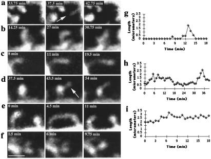
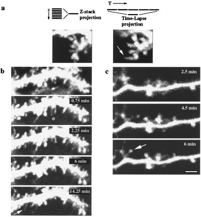
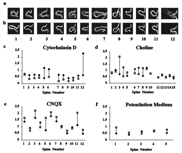
The function of dendritic spines, postsynaptic sites of excitatory input in the mammalian central nervous system (CNS), is still not well understood. Although changes in spine morphology may mediate synaptic plasticity, the extent of basal spine motility and its regulation and function remains controversial. We investigated spine motility in three principal neurons of the mouse CNS: cerebellar Purkinje cells, and cortical and hippocampal pyramidal neurons. Motility was assayed with time-lapse imaging by using two-photon microscopy of green fluorescent protein-labeled neurons in acute and cultured slices. In all three cell types, dendritic protrusions (filopodia and spines) were highly dynamic, exhibiting a diversity of morphological rearrangements over short (<1-min) time courses. The incidence of spine motility declined during postnatal maturation, but dynamic changes were still apparent in many spines in late-postnatal neurons. Although blockade or induction of neuronal activity did not affect spine motility, disruption of actin polymerization did. We hypothesize that this basal motility of dendritic protrusions is intrinsic to the neuron and underlies the heightened plasticity found in developing CNS.


Figures

Figure 1
Figure 1
GFP-transfected cells in slices have normal morphology and physiology. GFP-labeled Purkinje cells from P 10 + 2 div (a and b) sagittal or (c) frontal slices. Note labeled parallel fiber (arrow) in the frontal slice (c). (d and e) GFP-labeled hippocampal (P0 + 11 div) and (f and g) cortical (P1 + 22 div) pyramidal neurons. Individual dendritic spines on (b) Purkinje, (e) hippocampal, and (g) pyramidal neurons are clearly resolved at high magnification. (h) Whole-cell recording of action potentials elicited from a GFP-labeled cortical pyramidal neuron (10-hr acute slice) by injection of a depolarizing current. Bar = 50 μm in a, c, d, and f; 5 μm in b, e, and g.
Figure 2
Figure 2
Dendritic spines exhibit different types of morphological rearrangements. (a) A dendritic filopodium (arrow) appears and disappears (P). (b) Amorphous changes in the spine head (“morphing;” P). (c) Elongation of a spine (C). (d) Emergence of a filopodium (arrow) from a spine head (P). (e) Transient “touching” of neighboring spines (C). (f) Merging of split spine heads (P). (g) Length measurements of a transient dendritic spine, (h) a spine that elongates and retracts, and (i) a spine that elongates and remains elongated. Cellular origin of spines is denoted by: P, Purkinje; C, cortical pyramidal cells. Bar = 2 μm.
Figure 3
Figure 3
Spine motility does not result from focal-plane shifts, deafferentation, or slice-culture artifacts. (a Left) Projection of an “extended” 7-μm Z-stack (Purkinje cell, P10 + 2 div) spanning a volume above and below the plane of interest, collected before time-lapse imaging. (Right) Projection of several images from a time-lapse sequence into a single image. Note how the elongated spine that appears in the time-lapse projection (arrow) is not visible in the original “extended” Z-stack projection. (b) Spine motility in the frontal slices (Purkinje cell, P10 + 2 div), demonstrating retraction (∗) and appearance (arrow) of filopodia. (Top) A 7-μm Z-stack projection. (c) Time-lapse images from a cortical pyramidal neuron from an acute (10-hr) slice showing the appearance of a new spine (arrow). Bar = 2 μm.
Figure 4
Figure 4
Developmental regulation of spine motility. (a) A histogram of the percent motility at two developmental stages in Purkinje cells. Note how motility is reduced from 73.1 ± 11.8% at P10 + 2 div (n = 14) to 45 ± 14.9% at P22 + 2 div (n = 7); Mann–Whitney U test; P < 0.001). Developmental changes in motility in (b) acute and cultured cortical or (c) cultured hippocampal neurons. Age is computed as the sum of postnatal age and days in vitro (cultured slices were made at P0–1). The line represents the best fit to data from cultured cortical neurons (regression ANOVA F < 0.0006).
Figure 5
Figure 5
Spine motility is regulated by actin polymerization but is not affected by blocking or inducing activity. Superimposed outlines of spines (labeled 1–6 for Purkinje and 7–16 for cortical pyramidal neurons) from time-lapse sequences before a and after b application of Cytochalasin D. Motility indexes of the spines demonstrate that Cytochalasin D (c) reduces spine motility (Mann–Whitney U test, P = 0.003). (d)Lack of effect of choline on the motility indexes of Purkinje cells (spines 1–10) and cortical pyramidal neuron (spines 11–15). (e) Lack of effect of blocking AMPA receptors on motility (Purkinje cell). (f) Lack of effect of stimulating neuronal activity with potentiation medium (hippocampal pyramidal neuron). P > 0.5 for d–f.

References

    1. Cajal R Y. Rev Trimest Histol Norm Pat. 1888;1:1–10.
    1. Gray E G. J Anat. 1959;83:420–433. - PMC - PubMed
    1. Guthrie P B, Segal M, Kater S B. Nature (London) 1991;354:76–80. - PubMed
    1. Muller W, Connor J A. Nature (London) 1991;354:73–76. - PubMed
    1. Yuste R, Denk W. Nature (London) 1995;375:682–684. - PubMed

Publication types

Substances

LinkOut - more resources