. 1999 Nov 9;96(23):13450-5.
doi: 10.1073/pnas.96.23.13450.

Axon pathology in Parkinson's disease and Lewy body dementia hippocampus contains alpha-, beta-, and gamma-synuclein

Affiliations

Axon pathology in Parkinson's disease and Lewy body dementia hippocampus contains alpha-, beta-, and gamma-synuclein

J E Galvin et al. Proc Natl Acad Sci U S A. .

Abstract

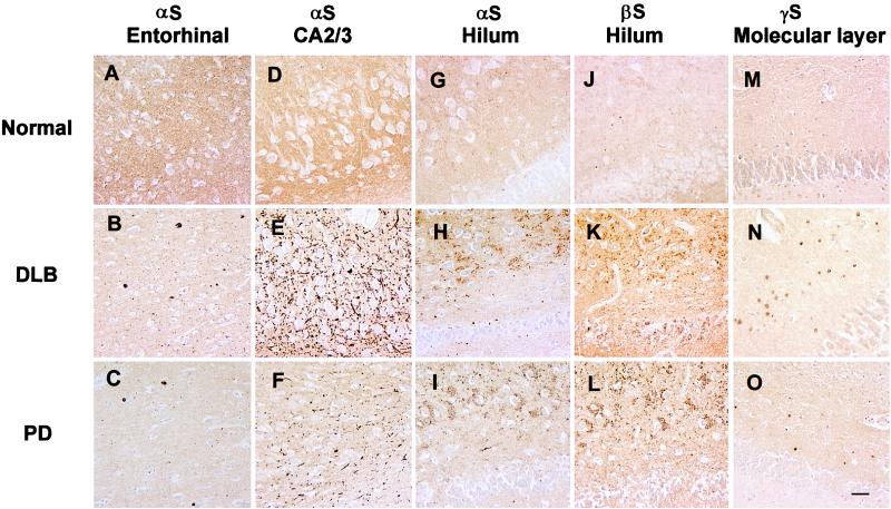
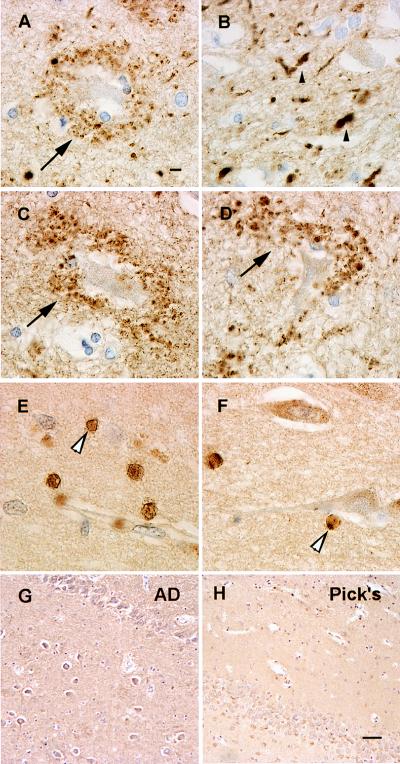
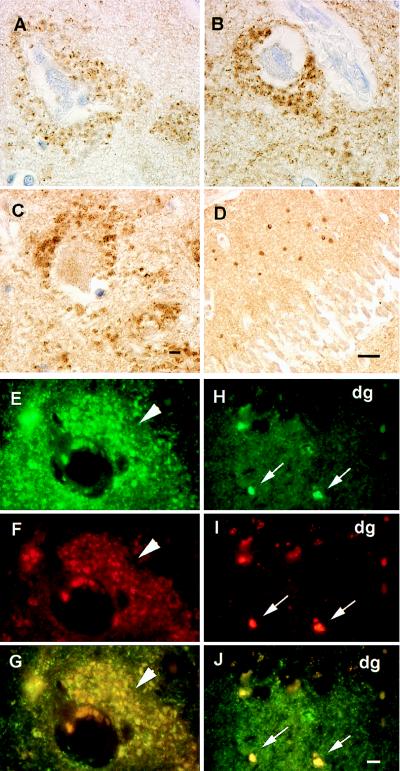
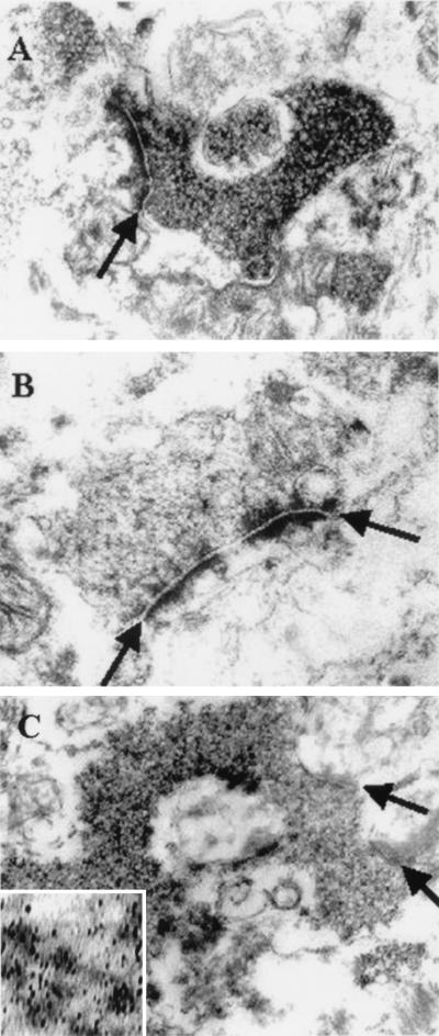
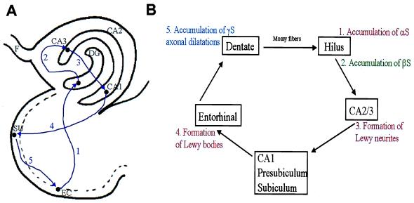
Pathogenic alpha-synuclein (alphaS) gene mutations occur in rare familial Parkinson's disease (PD) kindreds, and wild-type alphaS is a major component of Lewy bodies (LBs) in sporadic PD, dementia with LBs (DLB), and the LB variant of Alzheimer's disease, but beta-synuclein (betaS) and gamma-synuclein (gammaS) have not yet been implicated in neurological disorders. Here we show that in PD and DLB, but not normal brains, antibodies to alphaS and betaS reveal novel presynaptic axon terminal pathology in the hippocampal dentate, hilar, and CA2/3 regions, whereas antibodies to gammaS detect previously unrecognized axonal spheroid-like lesions in the hippocampal dentate molecular layer. The aggregation of other synaptic proteins and synaptic vesicle-like structures in the alphaS- and betaS-labeled hilar dystrophic neurites suggests that synaptic dysfunction may result from these lesions. Our findings broaden the concept of neurodegenerative "synucleinopathies" by implicating betaS and gammaS, in addition to alphaS, in the onset/progression of PD and DLB.


Figures

Figure 1
Figure 1
This series of low-power images depicts αS, βS, and γS immunoreactivity in hippocampus and entorhinal cortex of normal control (Top), DLB (Middle), and PD (Bottom) brains. The first column demonstrates the normal αS neuropil staining pattern (A) compared with the DLB (B) and PD (C) entorhinal cortex with αS-positive LBs using the LB509 antibody to αS. The second column shows the normal αS neuropil staining pattern (D) and dystrophic Lewy neurites in the CA2/3 region of hippocampus in the DLB case (E) and to a lesser extent in PD (F) by using LB509. The third column shows the normal αS neuropil staining pattern (G) and degenerate mossy fiber terminals around hilar neurons in DLB (H) and PD (I) by using LB509. The fourth column shows the normal αS neuropil staining pattern (J) as well as degenerate mossy fibers in DLB (K) and PD (L) by using the Syn207 antibody to βS. The final column shows the normal αS neuropil staining pattern (M) and depicts degenerate terminals in the stratum moleculare of the dentate gyrus in DLB (N) and PD (O) by using antisera 20 to γS. All panels are at the same magnification, and the scale bar in O = 20 μm.
Figure 2
Figure 2
This series of higher-power images depicts degenerate terminals labeled with antibodies to αS, βS, and γS in PD (A, G, and E) and DLB (B, D, and F) compared with AD (G) and Pick’s disease (H) where these lesions are not seen. (A) Degenerate mossy fiber terminals surrounding a hilar neuron with LB509 (arrow). (B) Lewy neurites and degenerate terminals on CA2/3 neurons (small arrowhead) with LB509. (C and D) Degenerating mossy fibers terminating on hilar neurons (arrow) with Syn207. (E) A high-power view of the γS immunoreactive terminals (large arrowheads) in the stratum moleculare of the dentate gyrus. (F) γS pathology in the CA1 region of the hippocampus (large arrowhead). (G) The absence of βS hilar pathology in AD. (H) Absence of γS pathology in Pick’s disease. A-F are at the same magnification (scale bar in A = 5 μm), and G and H are at the same magnification (scale bar in H = 20 μm).
Figure 3
Figure 3
Shown are degenerating mossy fiber terminals in PD and DLB cases with the presynaptic proteins synapsin, synaptophysin, and synaptobrevin. Degenerate mossy fiber terminals are labeled with antibodies against synapsin (A), synaptophysin (B), and synaptobrevin (C) identical to pathology seen with antibodies to αS and βS. (D) Degenerate terminals in the stratum moleculare stained by antibody against synaptobrevin similar to that seen with antisera 20 (γS). (EG) Double-label immunofluorescence of αS (green channel only in E) and synaptophysin (red channel only in F) terminals surrounding a hilar neuron (arrowheads), which colocalize to the same profiles in G (yellow immunofluorescence). (HJ) Double label immunofluorescence of γS (green channel only in H), synaptobrevin (red channel only in I), and their colocalization in J (yellow immunofluorescence identified by small arrows) in the molecular layer of the dentate gyrus (dg). AC and EJ are at the same magnification (scale bars in C and J = 5 μm). In D the scale bar = 20 μm.
Figure 4
Figure 4
The electron microscopic appearance of degenerate mossy fiber terminals. (A) The accumulation of αS immunoreactive vesicles in the presynaptic terminal of a mossy fiber rosette. (B) A control of a similar representative view without diaminobenzidine. (C) The αS immunoreactive vesicles demonstrated with silver enhancement technique. (Inset) A higher magnification of the αS immunoreactive synaptic vesicle. Arrows demonstrate synaptic clefts. Magnifications: ×45,000 for A–C; ×96,000 for Inset in C.
Figure 5
Figure 5
Perforant pathway and synuclein pathology. (A) The components of the hippocampal perforant pathway that play a critical role in behavior and memory. Neurons in entorhinal cortex send information in a unidirectional manner to dendrites in the molecular layer of neurons dentate gyrus (at 1), followed by hilar and CA2/3 neurons (at 2), and then to neurons in CA1 and presubicular (at 3) regions of the hippocampus. CA1 neurons then project back to the subiculum (at 4) and then to the entorhinal cortex (at 5) to complete this loop of interconnected neurons. (B) The perforant pathway in schematic form to illustrate the involvement of αS, βS, and γS pathology throughout this critical pathway in the PD and DLB hippocampus where synaptic transmission might be impaired by one or more synuclein lesions. EC, entorhinal cortex; SU, subiculum; DG, dentate gyrus; F, fornix.

References

    1. Ueda K, Fukushima H, Maliah E, Xia Y, Iwai A, Yoshimoto M, Otero D A, Kondo J, Ihara Y, Saitoh T. Proc Natl Acad Sci USA. 1993;90:11282–11286. - PMC - PubMed
    1. Jakes R, Spillantini M G, Goedert M. FEBS Lett. 1994;345:27–32. - PubMed
    1. Tobe T, Nakajo S, Tanaka A, Mitoya A, Omata K, Nakaya K, Tomita M, Nakmura Y. J Neurochem. 1992;59:1624–1629. - PubMed
    1. Nakajo S, Tsukada K, Omata K, Nakamura Y, Nakaya K. Eur J Biochem. 1993;217:1057–1063. - PubMed
    1. Ji H, Liu Y E, Jia T, Wang M, Liu J, Xiao G, Joseph B K, Rosen C, Shi Y E. Cancer Res. 1997;57:759–764. - PubMed

Publication types