. 1999 Nov 9;96(23):13474-9.
doi: 10.1073/pnas.96.23.13474.

INAF, a protein required for transient receptor potential Ca(2+) channel function

Affiliations

INAF, a protein required for transient receptor potential Ca(2+) channel function

C Li et al. Proc Natl Acad Sci U S A. .

Abstract

The trp gene of Drosophila encodes a subunit of a class of Ca(2+)-selective light-activated channels that carry the bulk of the phototransduction current. Transient receptor potential (TRP) homologs have been identified throughout animal phylogeny. In vertebrates, TRP-related channels have been suggested to mediate "store-operated Ca(2+) entry," which is important in Ca(2+) homeostasis in a wide variety of cell types. However, the mechanisms of activation and regulation of the TRP channel are not known. Here, we report on the Drosophila inaF gene, which encodes a highly eye-enriched protein, INAF, that appears to be required for TRP channel function. A null mutation in this gene significantly reduces the amount of the TRP protein and, in addition, specifically affects the TRP channel function so as to nearly shut down its activity. The inaF mutation also dramatically suppresses the severe degeneration caused by a constitutively active mutation in the trp gene. Although the reduction in the amount of the TRP protein may contribute to these phenotypes, several lines of evidence support the view that inaF mutations also more directly affect the TRP channel function, suggesting that the INAF protein may have a regulatory role in the channel function.


Figures
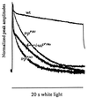
Figure 1
Figure 1
Comparison of the photoreceptor potentials of inaF and trp mutants. The receptor potentials elicited from inaFP106x, trpP301, trpP343, and wild-type photoreceptors are presented normalized to their peak amplitudes. White light stimuli (20 s) were used.
Figure 2
Figure 2
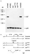
(A) Northern blot analysis of inaF mutants and controls. The six lanes were loaded with poly(A)+ RNA isolated from wild-type heads, wild-type bodies, eyes absent (eya) mutant heads, inaFP105p heads, inaFP106x heads, and revertant heads (lanes 1–6, respectively). Approximately 1.5 μg of poly(A)+ RNA was loaded in each lane. The probes were prepared from a 3.5-kb EcoRI fragment of the A23 genomic clone (B). Probes prepared from the inaF cDNA gave the same results. A single 3.2-kb transcript is detected only in wild-type and revertant heads (lanes 1 and 6). A small amount (<5% of wild-type amount) of the same transcript becomes visible in eya and inaFP105p heads (lanes 3 and 4), but not in wild-type bodies or inaF106x heads (lanes 2 and 5), upon overexposure of the blot (data not shown). The uniformity of RNA loading was monitored by reprobing the blot with rp49 cDNA (Bottom). (B) Restriction maps of inaF cDNA and of the corresponding genomic region in the A23 clone and three inaF mutants. The inverted triangle in the inaFP105p map identifies the site of P element insertion. The empty spaces to the right and left of the P insertion site in the inaFP106x and inaFP111x maps, respectively, represent the deletions caused by imprecise excision of the P element. In the cDNA map, the broken dotted line indicates the extent of the intron, and the open rectangle identifies the ORF. A composite genomic map at the top shows EcoRI sites (R) and the sizes of EcoRI fragments.
Figure 3
Figure 3
Immunodetection of the TRP protein. (A) Western blot analyses of null and near-null inaF and trp mutants and wild-type and revertant controls. The seven lanes were loaded with total protein prepared from wild-type heads, wild-type bodies, revertant heads, trpP301 heads, trpP343 heads, inaFP105p heads, and inaFP106x heads (lanes 1 to 7, respectively). The blot was probed with a monoclonal anti-TRP Ab (23). (B) Western blot analysis with retinal protein controls. The three lanes were loaded with total protein prepared from wild-type heads, inaFP106x heads, and trpP301 heads, and the blot was probed with, from top to bottom, anti-TRP, anti-INAD, anti-PLCβ, and anti-opsin Abs, respectively. (C) Subcellular localization of the TRP protein by immunofluorescence confocal microscopy. (Top) Confocal micrograph of the rhabdomeres of the inaFP106x compound eye labeled with a TRP antiserum and visualized with an FITC-conjugated secondary Ab. TRP-specific staining is found only in rhabdomeres. (Middle) Confocal micrograph of trpP301 rhabdomeres, processed exactly the same as for inaFP106x (see Methods). There is no TRP-specific staining. (Bottom) Confocal micrograph of exactly the same field and focal plane of trpP301 rhabdomeres as Middle, but showing staining of filamentous actin in rhabdomeres. The preparation was double-labeled with a TRP antiserum and phalloidin (see Methods). (D) Subcellular localization of control retinal proteins in inaF by confocal microscopy. Confocal micrographs of inaFP106x retinas labeled with anti-INAD (Top), anti-PLCβ (Middle), and anti-opsin (Bottom) Abs and visualized with an FITC-conjugated secondary Ab.
Figure 4
Figure 4
Electrophysiological analysis of inaFP106x in comparison to trp mutants. (A) Effects of inaFP106x mutation on the TRP (a) and TRPL (b) channel responses. (a) The receptor potentials recorded from the double mutants inaFP106x;trpl302, trpl302;trpP301, and trpl302;trpP343 are compared with that of trpl302. The peak amplitudes of the inaFP106x;trpl302, trpl302;trpP301, and trpl302 responses are, respectively, 13.1 ± 3.3, 10.3 ± 1.9, and 27 ± 3.6 mV. (b) The receptor potential recorded from trpP343 is compared with that from the double mutant inaFP106x;;trpP343. The wild-type response is as shown in Fig. 1. White light stimuli (20 s) were used throughout. (B) Refractory properties of the residual TRP responses of the double mutants, inaFP106x;trpl302 and trpl302;trpP301. Two successive stimuli (S1 and S2) separated by a 20-s interval (top trace) are presented to trpl302 (a), inaFP106x;trpl302 (b), and trpl302;trpP301 (c) following 2-min dark adaptation, and the resulting responses (R1 and R2) for each genotype are compared by superimposing. Control responses obtained from inaFP106x and trpP301 by using the same protocol are shown in d and e. The stimuli are white light of 2-s duration filtered with a 1-log-unit neutral density filter.
Figure 5
Figure 5
Suppression of the degeneration phenotype of TrpP365 by inaF. The time course of disappearance of the dpp, used as a measure of photoreceptor degeneration time course, is compared among TrpP365/+, trpP301, inaFP106x, and the double mutant, inaFP106x/inaFP106x;;TrpP365/+. All flies were raised under 12-hr-light/12-hr-dark illumination conditions.

Similar articles

Cited by

References

    1. Selinger Z, Minke B. Cold Spring Harbor Symp Quant Biol. 1988;53:333–341. - PubMed
    1. Bloomquist B B, Shortridge R D, Schneuwly S, Perdew M, Montell C, Steller H, Rubin G, Pak W L. Cell. 1988;54:723–733. - PubMed
    1. Toyoshima S, Matsumoto N, Wang P, Inoue H, Yoshioka T, Hotta Y, Osawa T. J Biol Chem. 1990;265:14842–14848. - PubMed
    1. Niemeyer B A, Suzuki E, Scott K, Jalink K, Zuker C S. Cell. 1996;85:651–659. - PubMed
    1. Hardie R C, Minke B. Neuron. 1992;8:613–651. - PubMed

Publication types

Associated data

LinkOut - more resources