. 1999 Nov 9;96(23):13486-90.
doi: 10.1073/pnas.96.23.13486.

Inosine stimulates extensive axon collateral growth in the rat corticospinal tract after injury

Affiliations

Inosine stimulates extensive axon collateral growth in the rat corticospinal tract after injury

L I Benowitz et al. Proc Natl Acad Sci U S A. .

Abstract

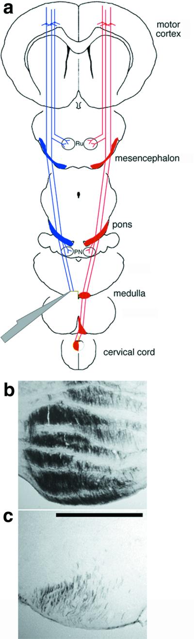
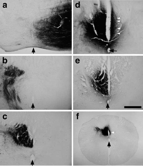
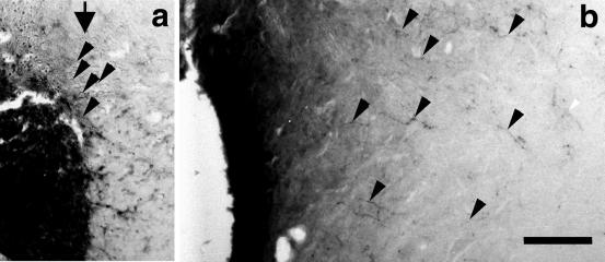
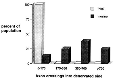
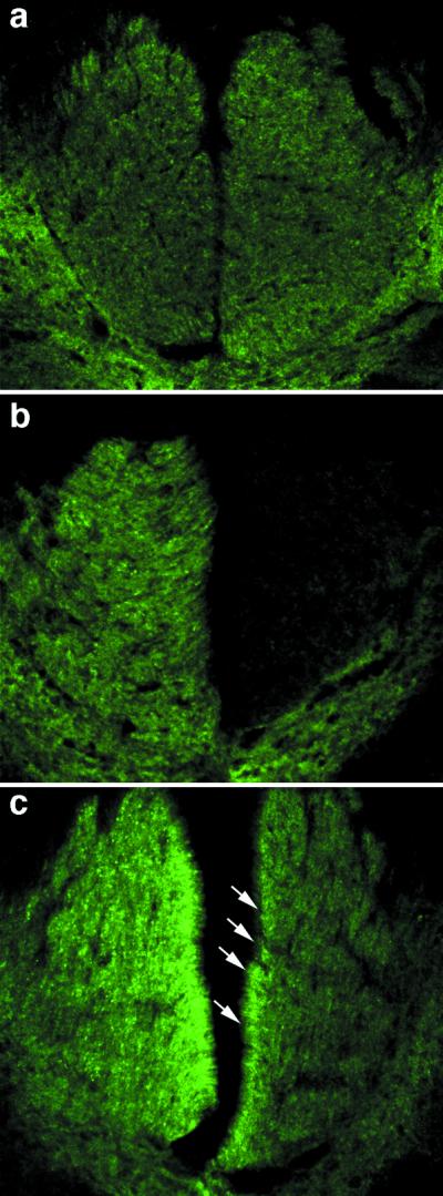
The purine nucleoside inosine has been shown to induce axon outgrowth from primary neurons in culture through a direct intracellular mechanism. For this study, we investigated the effects of inosine in vivo by examining whether it would stimulate axon growth after a unilateral transection of the corticospinal tract. Inosine applied with a minipump to the rat sensorimotor cortex stimulated intact pyramidal cells to undergo extensive sprouting of their axons into the denervated spinal cord white matter and adjacent neuropil. Axon growth was visualized by anterograde tracing with biotinylated dextran amine and by immunohistochemistry with antibodies to GAP-43. Thus, inosine, a naturally occurring metabolite without known side effects, might help to restore essential circuitry after injury to the central nervous system.


Figures

Figure 1
Figure 1
Unilateral transection of the CST. (a) CST axons arise from layer 5 pyramidal cells in the sensorimotor cortex, extend projections to the mesencephalic nucleus ruber (Ru) and pontine nuclei (PN), then assume a ventromedial position in the lower brainstem. In the caudal medulla, CST axons decussate and course in the dorsal funiculus of the contralateral spinal cord. Axons were transected via a ventral approach in the left medulla before the decussation. (b and c) Controls to verify the extent of surgery. Numerous fibers are seen in the left rostral medulla after left-sided BDA labeling (b); caudal to the transection, fewer than 10% of the original fibers remain. (c: bar = 500 μm.)
Figure 2
Figure 2
Inosine induces collateral sprouting in the adult rat CST. (a–d) Axon trajectories in a case in which we transected the CST in the left medulla (before the decussation), treated the right (nonaxotomized) sensorimotor cortex with inosine for 2 weeks, then traced fiber trajectories by injecting BDA into the right sensorimotor cortex. At more rostral levels, BDA-labeled axons remain strictly lateralized and are seen in the right ventral brainstem (a), left caudal medulla (b), and left dorsal funiculus (c). At the level of the cervical enlargement, however, numerous labeled axons also appear in the denervated (right-sided) dorsal funiculus (d; white arrowheads point to individual crossed fibers). (e) Controls treated with PBS instead of inosine after unilateral CST surgery show few crossed fibers. (Bar = 200 μm.) (f) A low-magnification image of the cervical cord from a case with left CST transection, inosine treatment in the right sensorimotor cortex, and BDA labeling in the right cortex. The white arrow points to dense axon staining on the denervated side. The midline is indicated in all sections with black arrows (pointing to the central canal in c–f).
Figure 3
Figure 3
Axon crossing and growth into the spinal cord gray matter. At the level of the cervical enlargement, labeled fibers (black arrowheads in a) are seen crossing the midline (black arrow). (b) In addition to numerous crossed fibers that remain close to the midline (left side of image), some crossed fibers course laterally into the denervated neuropil (arrowheads). (Bar = 100 μm.)
Figure 4
Figure 4
Axon crossings: frequency distributions for inosine- and PBS-treated groups. For each animal, we determined the mean number of crossed axons in the three coronal sections that showed the highest density of reinnervation. The x axis of the histogram shows the mean number of crossed axons, whereas the y axis represents the percentage of animals with axon numbers in each range. The distributions for numbers of crossed fibers show little overlap between the inosine-treated (n = 8; darkly shaded bars) and PBS-treated (n = 6; lightly shaded) groups.
Figure 5
Figure 5
Axon growth visualized by GAP-43 immunohistofluorescence. (a) At the midcervical level, modest levels of GAP-43 immunoreactivity appear bilaterally in the dorsal funiculi. (b) In control animals with unilateral CST transections and a 2-week infusion of PBS, GAP-43 immunoreactivity disappears from the denervated (right) dorsal funiculus, though a small number of fibers with increased staining appear in the intact side. (c) In animals with unilateral CST injury and inosine treatment, GAP-43 immunoreactivity intensifies on the intact (left) side and is also present in many fibers in the denervated (right) dorsal funiculus (white arrows show crossed fibers near the midline).

References

    1. Caroni P, Schwab M E. J Cell Biol. 1988;106:1281–1288. - PMC - PubMed
    1. Schnell L, Schwab M E. Nature (London) 1990;343:269–272. - PubMed
    1. McKerracher L, David S, Jackson D L, Kottis V, Dunn R J, Braun P E. Neuron. 1994;13:805–811. - PubMed
    1. Mukhopadhyay G, Doherty P, Walsh F S, Crocker P R, Filbin M T. Neuron. 1994;13:757–767. - PubMed
    1. Cheng H, Cao Y, Olson L. Science. 1996;273:510–513. - PubMed

Publication types

LinkOut - more resources