. 1999 Nov 9;96(23):13501-5.
doi: 10.1073/pnas.96.23.13501.

Semaphorin 3A growth cone collapse requires a sequence homologous to tarantula hanatoxin

Affiliations

Semaphorin 3A growth cone collapse requires a sequence homologous to tarantula hanatoxin

O Behar et al. Proc Natl Acad Sci U S A. .

Abstract

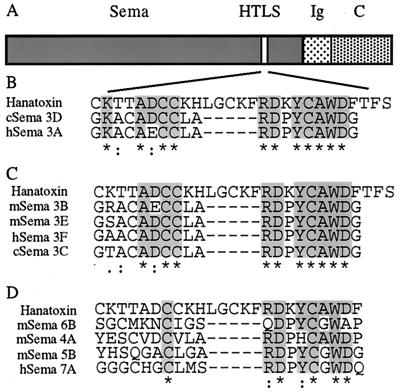
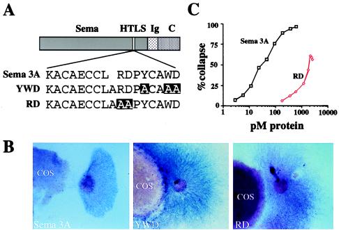
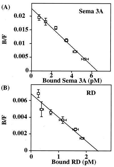
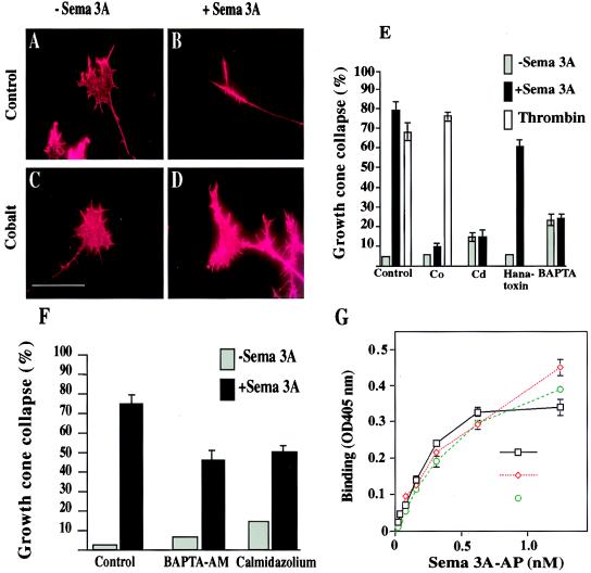
Axonal guidance is key to the formation of neuronal circuitry. Semaphorin 3A (Sema 3A; previously known as semaphorin III, semaphorin D, and collapsin-1), a secreted subtype of the semaphorin family, is an important axonal guidance molecule in vitro and in vivo. The molecular mechanisms of the repellent activity of semaphorins are, however, poorly understood. We have now found that the secreted semaphorins contain a short sequence of high homology to hanatoxin, a tarantula K(+) and Ca(2+) ion channel blocker. Point mutations in the hanatoxin-like sequence of Sema 3A reduce its capacity to repel embryonic dorsal root ganglion axons. Sema 3A growth cone collapse activity is inhibited by hanatoxin, general Ca(2+) channel blockers, a reduction in extracellular or intracellular Ca(2+), and a calmodulin inhibitor, but not by K(+) channel blockers. Our data support an important role for Ca(2+) in mediating the Sema 3A response and suggest that Sema 3A may produce its effects by causing the opening of Ca(2+) channels.


Figures

Figure 1
Figure 1
Primary sequence alignment of the semaphorin genes with hanatoxin. (A) Diagram of type III semaphorin genes (the secreted semaphorins). The three major domains, semaphorin domain (Sema), Ig-like domain (Ig), and carboxyl-terminal domain (C), are indicated. The site with sequence homology to hanatoxin (HTLS) is indicated. (B) Primary sequence alignment of the products of the two most homologous semaphorin genes, Sema 3A and Sema 3D, with hanatoxin (amino acids 9–35). (C) Sequence alignment of those secreted semaphorin gene products with less homology to hanatoxin. (D) Primary sequence alignment of four membrane-bound semaphorin. * indicates identity, : indicates high similarity, and . indicates low similarity.
Figure 2
Figure 2
Repulsion activity of Sema 3A HTLS mutants. (A) Schematic structure of Sema 3A protein showing wild-type and mutant sequences. (B) DRG explants cocultured with COS-7 cells expressing myc-Sema 3A, YWD mutant, or RD mutant were cultured for 40 hr. Note the strong repulsive effect in the wild-type transfected cells in comparison with the mutants. (Magnification, ×40.) (C) Dose–response curve, from one experiment, comparing RD-AP mutant growth cone collapse activity to Sema 3A-AP. Each concentration was repeated with at least 12 explants. EC50 for Sema 3A is 47 pM and for RD is 1466 pM.
Figure 3
Figure 3
Scatchard analysis of Sema 3A and RD mutant. Control PAE cells or PAE-NP-1 cells expressing neuropilin 1 (5 × 10⁴ cells) were treated with varying amounts of media containing Sema 3A-AP or RD-AP mutant. B/F, bound/free. Scatchard plots from one of the experiments for Sema 3A (A) and RD mutant (B) are presented. Kd value for Sema 3A-AP was 305 pM and for RD mutant was 505 pM. Bars indicate SEM for triplicates; where not obvious, the error bars are smaller than the symbols.
Figure 4
Figure 4
Extracellular Ca2+ influx and a Ca2+ channel are required for the collapse-inducing activity of Sema 3A. (A–E) E13 mouse embryo DRG explant cultures were incubated with or without various calcium channel blocking or depleting agents for 7 min prior to addition of control conditioned medium (A, C, and E) or Sema 3A-AP-conditioned medium (B, D, and E). (A and B) Control conditioned medium only; (C and D) with 2 mM CoCl2. (Scale bar, 50 μm.) (E) The relative responsiveness of DRG growth cones to Sema 3A-AP in the presence of 2 mM CoCl2, 200 μM CdCl2, 10 μM hanatoxin, and 1 mM BAPTA or thrombin (50 units/ml) in the presence or absence of 2 mM CoCl2. The percentage of collapsed growth cones with (black bars) or without (gray bars) Sema 3A-AP or with thrombin (white bars) is shown. (F) E13 mouse embryo DRG explant cultures were incubated with or without 2 μM BAPTA-AM and 0.08 μM calmidazolium chloride for 60 min prior to addition of control conditioned medium or Sema 3A-AP-conditioned media. The percentage of collapsed growth cones with (black bars) or without (gray bars) Sema 3A-AP in the presence of these reagents is presented. (G) Control PAE cells or PAE-NP-1 cells were treated with medium containing Sema 3A-AP with or without 2 mM CoCl2 or 200 μM CdCl2. Bars indicate SEM for triplicates. Where not obvious, the error bars are smaller than the symbols.

References

    1. Mark M D, Lohrum M, Puschel A W. Cell Tissue Res. 1997;290:299–306. - PubMed
    1. Luo Y, Raible D, Raper J A. Cell. 1993;75:217–227. - PubMed
    1. Kolodkin A L, Matthes D J, Goodman C S. Cell. 1993;75:1389–1399. - PubMed
    1. Behar O, Golden J A, Mashimo H, Schoen F J, Fishman M C. Nature (London) 1996;383:525–528. - PubMed
    1. Taniguchi M, Yuasa S, Fujisawa H, Naruse I, Saga S, Mishina M, Yagi T. Neuron. 1997;19:519–530. - PubMed

Publication types

LinkOut - more resources