. 1999 Nov 9;96(23):13518-23.
doi: 10.1073/pnas.96.23.13518.

Role of the Y5 neuropeptide Y receptor in limbic seizures

Affiliations

Role of the Y5 neuropeptide Y receptor in limbic seizures

D J Marsh et al. Proc Natl Acad Sci U S A. .

Abstract

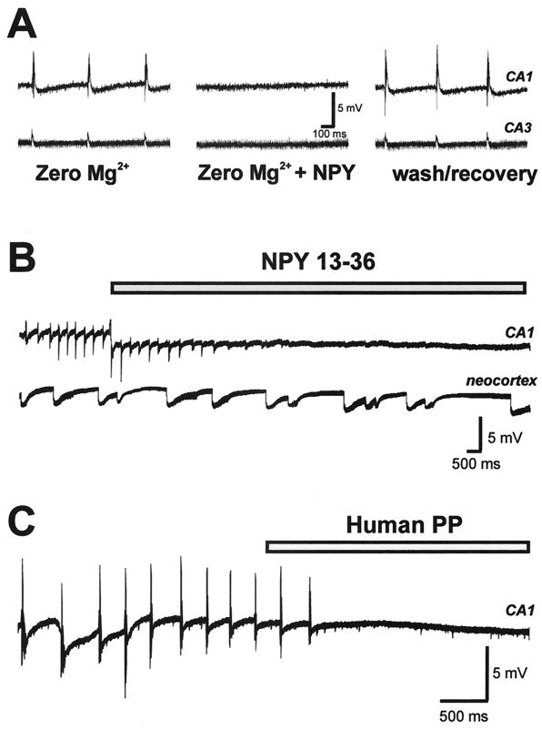
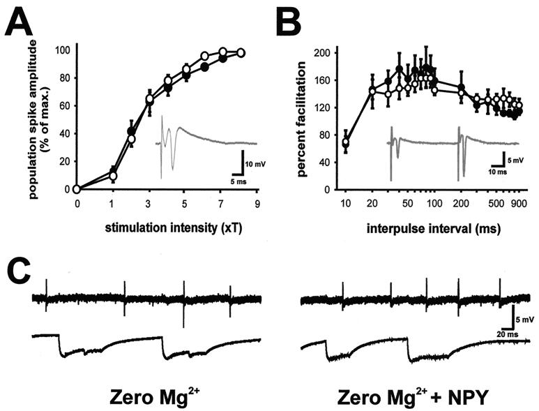
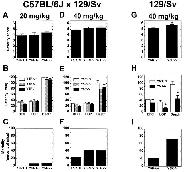
Neuropeptide Y (NPY) is an inhibitory neuromodulator expressed abundantly in the central nervous system that is suspected of being an endogenous antiepileptic agent that can control propagation of limbic seizures. Electrophysiological and pharmacological data suggest that these actions of NPY are mediated by G protein-coupled NPY Y2 and NPY Y5 receptors. To determine whether the NPY Y5 receptor (Y5R) is required for normal control of limbic seizures, we examined hippocampal function and responsiveness to kainic acid-induced seizures in Y5R-deficient (Y5R-/-) mice. We report that Y5R-/- mice do not exhibit spontaneous seizure-like activity; however, they are more sensitive to kainic acid-induced seizures. Electrophysiological examination of hippocampal slices from mutant mice revealed normal function, but the antiepileptic effects of exogenously applied NPY were absent. These data demonstrate that Y5R has an important role in mediating NPY's inhibitory actions in the mouse hippocampus and suggest a role for Y5R in the control of limbic seizures.


Figures

Figure 1
Figure 1
NPY blockade of spontaneous epileptiform burst discharge in hippocampal slices from wild-type mice. Epileptiform activity was induced by slice perfusion with bathing medium containing no magnesium. (A) Field recordings were obtained simultaneously from CA1 (Upper) and CA3 (Lower) pyramidal cell layers. After ≈45 min of perfusion with 0-Mg-ACSF, spontaneous burst discharges occurred in CA1 and CA3. This discharge pattern was abolished after a 10-min perfusion with 0-Mg-ACSF containing 1 μM human NPY. Recovery of burst activity was seen within 15 min after return to 0-Mg-ACSF. (B) Spontaneous epileptiform burst discharges occurring in CA1 were abolished by perfusion with 0-Mg-ACSF containing 1 μM human NPY13–36; burst-discharge activity occurring in neocortex (layer II–III) was unchanged. (C) Spontaneous epileptiform burst discharges occurring in CA1 were abolished by perfusion with 0-Mg-ACSF containing 1 μM human pancreatic polypeptide.
Figure 2
Figure 2
Synaptic function and epileptiform-discharge activity in hippocampal slices from Y5R−/− mice. (A) Input–output curves of population spike responses recorded in the CA1 pyramidal cell region (see Inset) to stimulation of the Schaffer collaterals. Threshold for stimulation was defined for each slice as the minimum current required to elicit a detectable population spike; the x axis shows stimulus intensity in terms of threshold multiples. Responses were normalized with respect to maximum population spike amplitude to allow averaging of responses from wild-type (○) and Y5R−/− (●) mice. (B) Plot of paired-pulse facilitation (amplitude of population spike response to second stimulus divided by amplitude of the response to first stimulus) in the CA1 pyramidal cell region (see Inset) for hippocampal slices from wild-type and Y5R−/− mice. Stimulation intensity was set at four times the threshold. (C) Spontaneous epileptiform burst-discharge activity in a hippocampal slice from a Y5R−/− mouse during perfusion with 0-Mg-ACSF (Left) and 20 min after perfusion with 0-Mg-ACSF containing 1 μM human NPY (Right). Field recordings were obtained from hippocampus (CA1 pyramidal cell layer, Upper) and neocortex (layer II–III, Lower).
Figure 3
Figure 3
Response of wild-type and mutant mice to peripherally administered KA. Female wild-type (Y5R+/+), heterozygous (Y5R+/−), and homozygous Y5R−/− littermate mice on a mixed C57BL/6J × 129/Sv (A–F) genetic background or female wild-type and Y5R−/− mice on an inbred 129/Sv (G–I) genetic background were injected (i.p.) with either 20 (A–C) or 40 (D–I) mg/kg KA and observed for 2 hr after injection. (A, D, and G) Motor seizure severity was rated on a scale from 0 to 6 as described in Materials and Methods (n = 16–35 per genotype; *, P < 0.001). (B, E, and H) Latency to loss of posture (LOP), bilateral forelimb clonus (BFC), and death in response to KA was measured in minutes for control and mutant mice (n = 9–35 per genotype; *, P < 0.004). (C, F, and I) Plot of the percent mortality of control and mutant mice after KA administration (n = 16–35 per genotype).

References

    1. Allen Y S, Adrian T E, Allen J M, Tatemoto K, Crow T J, Bloom S R, Polak J M. Science. 1983;221:877–879. - PubMed
    1. Chronwall B M, DiMaggio D A, Massari V J, Pickel V M, Ruggiero D A, O’Donohue T L. Neuroscience. 1985;15:1159–1181. - PubMed
    1. Dumont Y, Martel J C, Fournier A, St.-Pierre S, Quirion R. Prog Neurobiol. 1992;38:125–167. - PubMed
    1. Morris B J. J Comp Neurol. 1989;290:358–368. - PubMed
    1. Gruber B, Greber S, Rupp E, Sperk G. Hippocampus. 1994;4:474–482. - PubMed