

Long-term expression of protein kinase C in adult mouse hearts improves postischemic recovery
- PMID: 10557356
- PMCID: PMC23983
- DOI: 10.1073/pnas.96.23.13536
Long-term expression of protein kinase C in adult mouse hearts improves postischemic recovery
Abstract
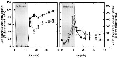
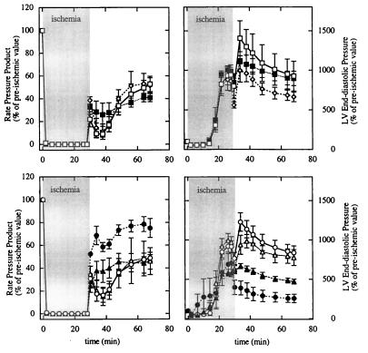
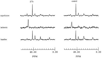
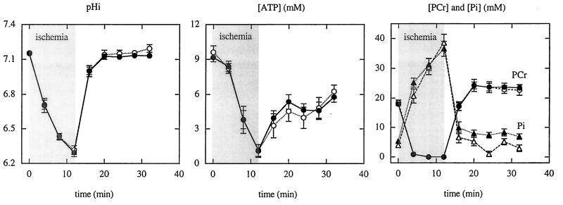
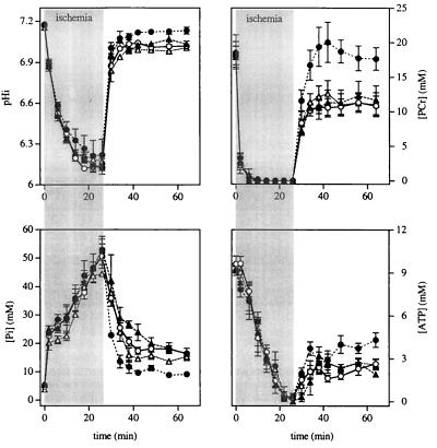
Activation of protein kinase C (PKC) protects the heart from ischemic injury; however, its mechanism of action is unknown, in part because no model for chronic activation of PKC has been available. To test whether chronic, mild elevation of PKC activity in adult mouse hearts results in myocardial protection during ischemia or reperfusion, hearts isolated from transgenic mice expressing a low level of activated PKCbeta throughout adulthood (beta-Tx) were compared with control hearts before ischemia, during 12 or 28 min of no-flow ischemia, and during reperfusion. Left-ventricular-developed pressure in isolated isovolumic hearts, normalized to heart weight, was similar in the two groups at baseline. However, recovery of contractile function was markedly improved in beta-Tx hearts after either 12 (97 +/- 3% vs. 69 +/- 4%) or 28 min of ischemia (76 +/- 8% vs. 48 +/- 3%). Chelerythrine, a PKC inhibitor, abolished the difference between the two groups, indicating that the beneficial effect was PKC-mediated. (31)P NMR spectroscopy was used to test whether modification of intracellular pH and/or preservation of high-energy phosphate levels during ischemia contributed to the cardioprotection in beta-Tx hearts. No difference in intracellular pH or high-energy phosphate levels was found between the beta-Tx and control hearts at baseline or during ischemia. Thus, long-term modest increase in PKC activity in adult mouse hearts did not alter baseline function but did lead to improved postischemic recovery. Furthermore, our results suggest that mechanisms other than reduced acidification and preservation of high-energy phosphate levels during ischemia contribute to the improved recovery.
Figures
References
-
- Puceat M, Brown J H. In: Protein Kinase C. Kuo J F, editor. Oxford: Oxford Univ. Press; 1994. pp. 249–268.
-
- Mitchell M B, Meng X, Ao L, Brown J M, Harken A H, Banerjee A. Circ Res. 1995;76:73–81. - PubMed
-
- Cohen M V, Downey J M. Annu Rev Med. 1996;47:21–29. - PubMed
-
- Liu Y, Tsuchida A, Cohen M V, Downey J M. J Mol Cell Cardiol. 1995;27:883–892. - PubMed
-
- Bugge E, Ytrehus K. Cardiovasc Res. 1996;32:920–929. - PubMed
Publication types
MeSH terms
Substances
Grants and funding
LinkOut - more resources
Full Text Sources
Molecular Biology Databases
