

doi: 10.1136/bmj.319.7220.1302.
Brain imaging: the NMR revolution. Interview by Clare Thompson
- PMID: 10559054
- PMCID: PMC1129079
- DOI: 10.1136/bmj.319.7220.1302
Item in Clipboard
Brain imaging: the NMR revolution. Interview by Clare Thompson
BMJ.
.
No abstract available
Figures
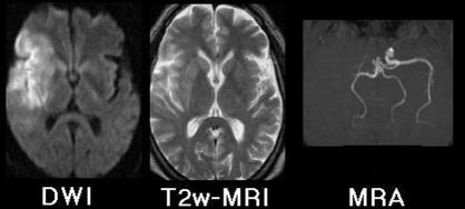
Magnetic resonance images 9.5 hours after onset of clinical stroke syndrome. Diffusion weighted imaging (DWI) shows hyperintense signal from physiologically damaged tissue surrounding right sylvian fissure—conventional T2 weighted imaging shows only subtle changes in same region. Magnetic resonance angiography (MRA) shows flow void in right internal carotid and middle cerebral arteries
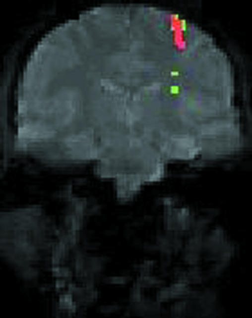
Functional magnetic resonance image of normal subject performing finger movements (conventional coronal magnetic resonance image in grey). Coloured regions identify tissue in which blood flow increases to meet extra metabolic requirements of task
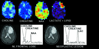
(Top) Spectroscopic image of patient with glioblastoma multiforme. Colour images display topographical distribution of intensities of NMR signals from several cerebral metabolites including total choline, total creatine, N-acetylaspartate, lactate, and lipid. (Bottom) Tissue section examined displayed by conventional magnetic resonance imaging. Metabolite signals can be displayed as spectra from volumes (white squares) referenced to magnetic resonance image (graphs). Compared with right frontal lobe, neoplastic tissue in left frontal lobe shows increased choline, lactate, and lipid signals but decreased N-acetylaspartate signals
Similar articles
-
Brain imaging in psychiatry: what has it done for the patient?Hosp Med. 2002 Jun;63(6):326-7. doi: 10.12968/hosp.2002.63.6.2000. Hosp Med. 2002. PMID: 12096659 No abstract available.
-
[Functional brain imaging in psychiatry].Duodecim. 2000;116(4):409-15. Duodecim. 2000. PMID: 11787093 Review. Finnish. No abstract available.
-
Clinical applications of neuroimaging in psychiatry.Magn Reson Imaging Clin N Am. 1998 Feb;6(1):155-64. Magn Reson Imaging Clin N Am. 1998. PMID: 9449746 Review.
-
Pretherapeutic functional magnetic resonance imaging in children.Neuroimaging Clin N Am. 2014 Nov;24(4):639-53. doi: 10.1016/j.nic.2014.07.002. Epub 2014 Nov 1. Neuroimaging Clin N Am. 2014. PMID: 25441505 Review.
-
Very like a fish.BMJ. 2011 Aug 3;343:d4918. doi: 10.1136/bmj.d4918. BMJ. 2011. PMID: 21816746 No abstract available.
Cited by
-
Purinergic system dysfunction in mood disorders: a key target for developing improved therapeutics.Prog Neuropsychopharmacol Biol Psychiatry. 2015 Mar 3;57:117-31. doi: 10.1016/j.pnpbp.2014.10.016. Epub 2014 Nov 7. Prog Neuropsychopharmacol Biol Psychiatry. 2015. PMID: 25445063 Free PMC article.
References
-
- Stark DD, Bradley WG. Magnetic resonance imaging. St Louis: Mosby; 1992.
-
- Grant DM, Harris RK. Encyclopedia of NMR. Chichester: Wiley; 1996.
-
- Budinger TF. MR safety: past, present, and future from a historical perspective. Magn Reson Imaging Clin North Am. 1998;6:701–714. - PubMed
-
- Le Bihan D. Diffusion and perfusion magnetic resonance imaging: applications to functional MRI. New York: Raven; 1995.
-
- Warach S, Gaa J, Siewert B, Wielopolski P, Edelman RR. Acute human stroke studied by whole brain echo planar diffusion-weighted magnetic resonance imaging. Ann Neurol. 1995;37:231–241. - PubMed
Publication types
MeSH terms
Grants and funding
LinkOut - more resources
Full Text Sources
Medical