

Characterization of a periplasmic ATP-binding cassette iron import system of Brachyspira (Serpulina) hyodysenteriae
- PMID: 10559160
- PMCID: PMC94169
- DOI: 10.1128/JB.181.22.6948-6957.1999
Characterization of a periplasmic ATP-binding cassette iron import system of Brachyspira (Serpulina) hyodysenteriae
Abstract
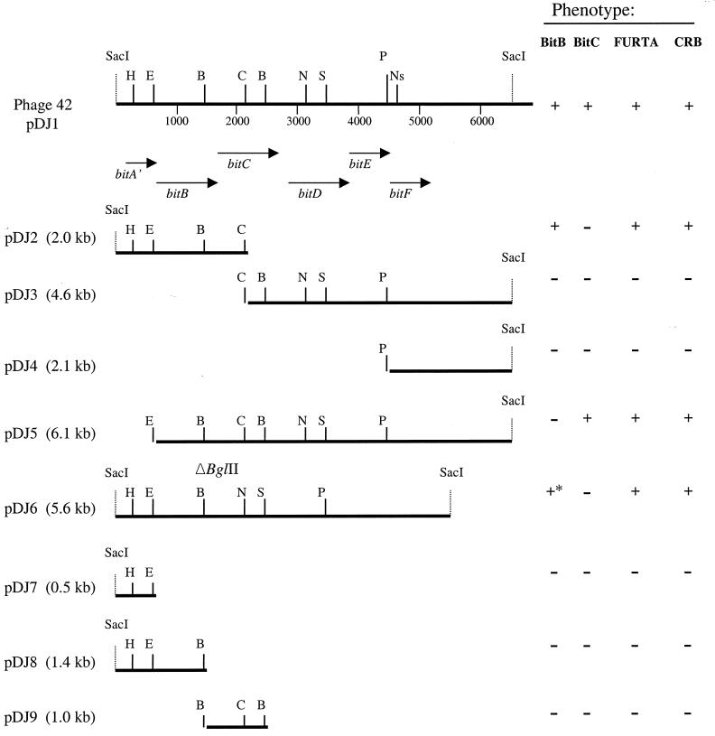
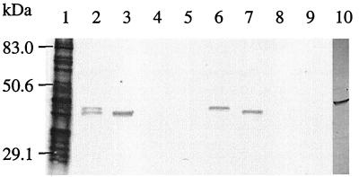
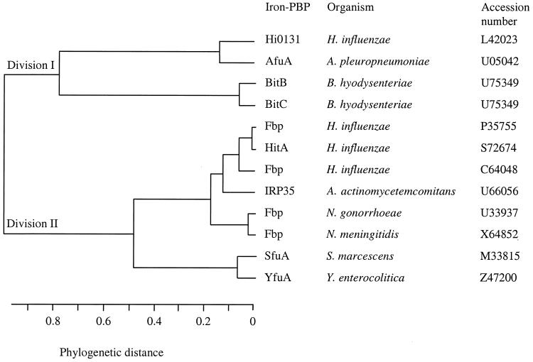
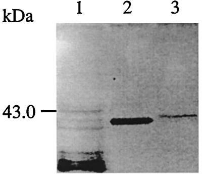
The nucleotide sequence of the pathogenic spirochete Brachyspira hyodysenteriae bit (for "Brachyspira iron transport") genomic region has been determined. The bit region is likely to encode an iron ATP-binding cassette transport system with some homology to those encountered in gram-negative bacteria. Six open reading frames oriented in the same direction and physically linked have been identified. This system possesses a protein containing ATP-binding motifs (BitD), two hydrophobic cytoplasmic membrane permeases (BitE and BitF), and at least three lipoproteins (BitA, BitB, and BitC) with homology to iron periplasmic binding proteins. These periplasmic binding proteins exhibit lipoprotein features. They are labeled by [(3)H]palmitate when tested in recombinant Escherichia coli, and their signal peptides are typical for substrates of the type II secretory peptidase. The FURTA system and Congo red assay indicate that BitB and BitC are involved in iron binding. The Bit system is detected only in B. hyodysenteriae and is absent from B. innocens and B. pilosicoli.
Figures
Similar articles
-
Cloning and DNA sequence analysis of an immunogenic glucose-galactose MglB lipoprotein homologue from Brachyspira pilosicoli, the agent of colonic spirochetosis.Infect Immun. 2000 Aug;68(8):4559-65. doi: 10.1128/IAI.68.8.4559-4565.2000. Infect Immun. 2000. PMID: 10899855 Free PMC article.
-
Identification of the gene encoding BmpB, a 30 kDa outer envelope lipoprotein of Brachyspira (Serpulina) hyodysenteriae, and immunogenicity of recombinant BmpB in mice and pigs.Vet Microbiol. 2000 Oct 1;76(3):245-57. doi: 10.1016/s0378-1135(00)00244-3. Vet Microbiol. 2000. PMID: 10973699
-
Unification of the genera Serpulina and Brachyspira, and proposals of Brachyspira hyodysenteriae Comb. Nov., Brachyspira innocens Comb. Nov. and Brachyspira pilosicoli Comb. Nov.Microbiol Immunol. 1997;41(6):445-52. doi: 10.1111/j.1348-0421.1997.tb01877.x. Microbiol Immunol. 1997. PMID: 9251055
-
Fate of ferrisiderophores after import across bacterial outer membranes: different iron release strategies are observed in the cytoplasm or periplasm depending on the siderophore pathways.Amino Acids. 2013 May;44(5):1267-77. doi: 10.1007/s00726-013-1468-2. Epub 2013 Feb 27. Amino Acids. 2013. PMID: 23443998 Review.
-
ABC transporters involved in the biogenesis of the outer membrane in gram-negative bacteria.Biosci Biotechnol Biochem. 2011;75(6):1044-54. doi: 10.1271/bbb.110115. Epub 2011 Jun 13. Biosci Biotechnol Biochem. 2011. PMID: 21670534 Review.
Cited by
-
Phylogenetic diversity, antimicrobial susceptibility and virulence gene profiles of Brachyspira hyodysenteriae isolates from pigs in Germany.PLoS One. 2018 Jan 11;13(1):e0190928. doi: 10.1371/journal.pone.0190928. eCollection 2018. PLoS One. 2018. PMID: 29324785 Free PMC article.
-
Genomic insights into the population structure, antimicrobial resistance, and virulence of Brachyspira hyodysenteriae from diverse geographical regions.Microbiol Spectr. 2025 Jun 3;13(6):e0338624. doi: 10.1128/spectrum.03386-24. Epub 2025 Apr 24. Microbiol Spectr. 2025. PMID: 40272172 Free PMC article.
-
The Exposed Proteomes of Brachyspira hyodysenteriae and B. pilosicoli.Front Microbiol. 2016 Jul 21;7:1103. doi: 10.3389/fmicb.2016.01103. eCollection 2016. Front Microbiol. 2016. PMID: 27493641 Free PMC article.
-
Comparison of Brachyspira hyodysenteriae Isolates Recovered from Pigs in Apparently Healthy Multiplier Herds with Isolates from Herds with Swine Dysentery.PLoS One. 2016 Aug 4;11(8):e0160362. doi: 10.1371/journal.pone.0160362. eCollection 2016. PLoS One. 2016. PMID: 27489956 Free PMC article.
-
The S. aureus 4-oxalocrotonate tautomerase SAR1376 enhances immune responses when fused to several antigens.Sci Rep. 2017 May 11;7(1):1745. doi: 10.1038/s41598-017-01421-z. Sci Rep. 2017. PMID: 28496136 Free PMC article.
References
-
- Adhikari P, Kirby S D, Nowalk A J, Veraldi K L, Schyvers A B, Mietzner T A. Biochemical characterization of a Haemophilus influenzae periplasmic iron transport operon. J Biol Chem. 1995;270:25142–25149. - PubMed
-
- Ames G F. Bacterial periplasmic transport systems: structure, mechanism, and evolution. Annu Rev Biochem. 1986;55:397–425. - PubMed
-
- Ames G F-L. Energetics of periplasmic transport systems. In: Krulwich T A, editor. Bacterial energetics. New York, N.Y: Academic Press; 1990. pp. 225–245.
Publication types
MeSH terms
Substances
Associated data
- Actions
LinkOut - more resources
Full Text Sources
Other Literature Sources
Medical
