. 1999 Nov;181(22):7028-33.
doi: 10.1128/JB.181.22.7028-7033.1999.

Transcriptional control of the low-temperature-inducible des gene, encoding the delta5 desaturase of Bacillus subtilis

Affiliations

Transcriptional control of the low-temperature-inducible des gene, encoding the delta5 desaturase of Bacillus subtilis

P S Aguilar et al. J Bacteriol. 1999 Nov.

Abstract

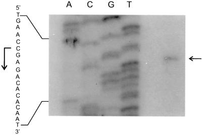
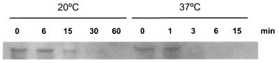
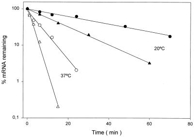
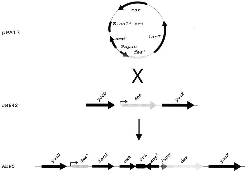
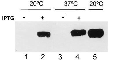
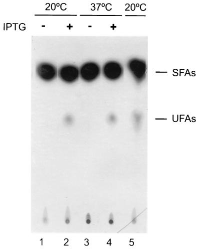
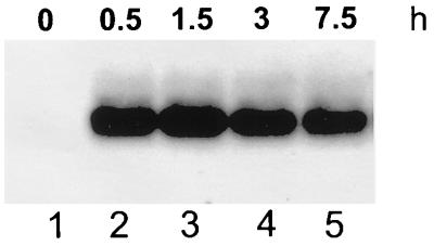
The Bacillus subtilis des gene encodes the cold-inducible Delta5 lipid desaturase involved in the formation of unsaturated fatty acids from saturated phospholipid precursors. Here, we describe the expression pattern of the des gene in response to a temperature downshift from 37 to 20 degrees C. We found that the synthesis of des mRNA is undetectable at 37 degrees C but dramatically induced upon the temperature downshift. Decay characteristics of the des transcript as well as the in vivo decay of B. subtilis bulk mRNA were investigated. The results showed that the stability of the des transcript as well as of bulk mRNA lasted substantially longer at 20 degrees C than at 37 degrees C. Functional expression of des at 37 degrees C was achieved by exchanging its promoter with the non-cold shock spac promoter. These data provide the first direct evidence that temperature-mediated control of transcription is the major mechanism regulating the mRNA levels of the B. subtilis desaturase. The present results also demonstrate that the only component of the desaturation system regulated by temperature is the desaturase enzyme.


Figures

FIG. 1
FIG. 1
des mRNA production before and after cold shock at 20°C and in the absence or presence of 0.2 mg of chloramphenicol per ml. (A) Northern blot analysis with formaldehyde-agarose gels was carried out as described in Materials and Methods. Total RNA was isolated from strain JH642 grown until mid-exponential phase at 37°C (lane 1), from cells shifted from 37 to 20°C at different times (lanes 2 to 8) and from cells grown continuously at 20°C (lane 9). Each lane contains 8 μg of total RNA. RNA blots were probed with a des-specific probe. After autoradiography, blots were stripped and reprobed with a B. subtilis 23S rRNA-specific probe. Autoradiographs of the resulting blots are shown. (B) Graph of the results shown in panel A. The densities of the bands corresponding to des mRNA and 23S rRNA for each lane were measured by a densitometer. The ratio of the intensities of 23S and des was used for the plot. (C) Northern blot analysis was performed as described for panel A. Cells were grown at 37°C until mid-exponential phase (lane 1) and then treated with chloramphenicol (200 μg/ml) for 15 min (lane 3) or then shifted to 20°C for 45 min in the absence (lane 2) or presence (lane 4) of chloramphenicol (200 μg/ml).
FIG. 2
FIG. 2
Primer extension analysis of the transcription start site. The autoradiogram shows a primer extension experiment performed with RNA extracted from strain JH642 incubated at 20°C for 45 min after shifting from 37°C. Lanes A, C, G, and T show sequencing reactions performed on des with the primer extension oligonucleotide. The horizontal arrow indicates the hybridizing extension product corresponding to the C residue taken to be the start of transcription. The vertical arrow indicates the direction of transcription.
FIG. 3
FIG. 3
Northern blot analysis of des mRNA decay after rifampin addition. Total RNA (5 μg per lane) from strain JH642 was separated on a denaturing 5% polyacrylamide gel. Cells were grown at 37°C until mid-exponential phase, des mRNA synthesis was induced by transferring the culture to 20°C for 45 min, rifampin (final concentration, 500 μg/ml) and nalidixic acid (final concentration, 20 μg/ml) were added, and the cells were kept at 20°C or transferred to 37°C. Total RNA from aliquots of 25 ml was extracted after the addition of the antibiotics (times shown indicate minutes elapsed following addition of antibiotics).
FIG. 4
FIG. 4
Stabilities of des and bulk mRNA. The des mRNA levels at 37°C (open triangles) or 20°C (filled triangles) were determined by quantifying the radioactivity of the bands of the Northern blot shown in Fig. 3 and from dot blots of RNA hybridized with a radiolabeled probe specific for des, as described in Materials and Methods. The samples for dot blot analysis were taken at the same times as those for the Northern analysis, as indicated in the legend of Fig. 3. Each value is the average of the results of two independent experiments. The bulk RNA decay was measured as described in Materials and Methods. Samples at 37°C (open circles) were taken at 0, 1, 3, 6, 12, and 24 min after inhibition of RNA synthesis. Samples at 20°C (filled circles) were taken at 0, 6, 12, 24, 48, and 70 min after inhibition of RNA synthesis.
FIG. 5
FIG. 5
Construction of AKP5 strain with des under spac promoter control. Plasmid pPA13, which contains the 28-nucleotide-long 5′ UTR and the first 147 codons of the des gene downstream of the spac promoter, was used to transform wild-type strain JH642 to Cmr, yielding strain AKP5. Campbell insertion of this plasmid places the des gene under the control of the spac promoter.
FIG. 6
FIG. 6
des mRNA production under the control of the spac promoter. Total RNA (10 μg/ml) from strain AKP5 (lanes 1 to 4) or JH642 (lane 5) was separated on formaldehyde-agarose gels. Strain AKP5 was grown at 37°C to mid-exponential phase, and half of this culture was supplemented with 1 mM of IPTG. Treated (+) and untreated (−) cultures were further shifted to 20°C for 45 min (lanes 1 and 2) or maintained at 37°C for 15 min (lanes 3 and 4). Strain JH642 was grown at 37°C to mid-exponential phase and then shifted to 20°C for 45 min (lane 5).
FIG. 7
FIG. 7
Fatty acids synthesized by strain AKP5 at 37 and 20°C. Cultures of strain AKP5 (lanes 1 to 4) were grown to mid-exponential phase at 37°C. One half of this culture was supplemented with 1 mM of IPTG. Two milliliters of treated (+) and untreated (−) cultures was challenged with 10 μCi of [14C]acetate and further shifted to 20°C (lanes 1 and 2) or maintained at 37°C (lanes 3 and 4) for 12 h. The lipids were then extracted and transesterified, and the resulting methyl esters were separated into saturated fatty acid (SFA) and UFA fractions by chromatography on 20% silver nitrate impregnated silica gel thin-layer plates. The plates were developed at −17°C and autoradiographed for 5 days. The UFA synthesized by strain JH642 grown at 37°C and then shifted to 20°C in the presence of 10 μCi of [14C]acetate for 12 h is shown in lane 5. The samples in lanes 1 and 3 contained 11,000 cpm in the SFA fractions, while the UFA fractions contained only background levels of radioactivity. The samples in lanes 2 and 4 contained 10,000 and 1,300 cpm of radioactivity in the SFA and UFA fractions, respectively. The sample in lane 5 contained 9,000 and 1,350 cpm of radioactivity in the SFA and UFA fractions, respectively.
FIG. 8
FIG. 8
Pattern of spac-des expression at 20°C. Total RNA (10 μg/ml) from strain AKP5 was separated on formaldehyde-agarose gels. Cells were grown at 37°C to mid-exponential phase, supplemented with 1 mM IPTG, and then shifted to 20°C. Total RNA was extracted after the addition of IPTG (times shown indicate hours elapsed following addition of IPTG).

Similar articles

Cited by

References

    1. Aguilar P S, Cronan J E, Jr, de Mendoza D. A Bacillus subtilis gene induced by cold shock encodes a membrane phospholipid desaturase. J Bacteriol. 1998;180:2194–2200. - PMC - PubMed
    1. Aguilar, P. S., and D. de Mendoza. 1998. Unpublished results.
    1. Cronan J E, Jr, Rock C O. Biosynthesis of membrane lipids. In: Neidhardt F C, Curtiss III R, Ingraham J L, Lin E C C, Low K B, Magasanik B, Reznikoff W S, Riley M, Schaechter M, Umbarger H E, editors. Escherichia coli and Salmonella: cellular and molecular biology. 2nd ed. Vol. 1. Washington, D.C.: American Society for Microbiology; 1996. pp. 612–636.
    1. de Mendoza D, Grau R, Cronan J E., Jr . Biosynthesis and function of membrane lipids. In: Sonenshein A L, Hoch J A, Losick R, editors. Bacillus subtilis and other gram-positive bacteria: biochemistry, physiology, and molecular genetics. Washington, D.C.: American Society for Microbiology; 1993. pp. 411–421.
    1. Dowhan W. Molecular basis for membrane phospholipid diversity: why are there so many lipids? Annu Rev Biochem. 1997;66:199–232. - PubMed

Publication types

MeSH terms

LinkOut - more resources