. 1999 Nov;181(22):7043-51.
doi: 10.1128/JB.181.22.7043-7051.1999.

Functional regions of the Bacillus subtilis spore coat morphogenetic protein CotE

Affiliations

Functional regions of the Bacillus subtilis spore coat morphogenetic protein CotE

T Bauer et al. J Bacteriol. 1999 Nov.

Abstract

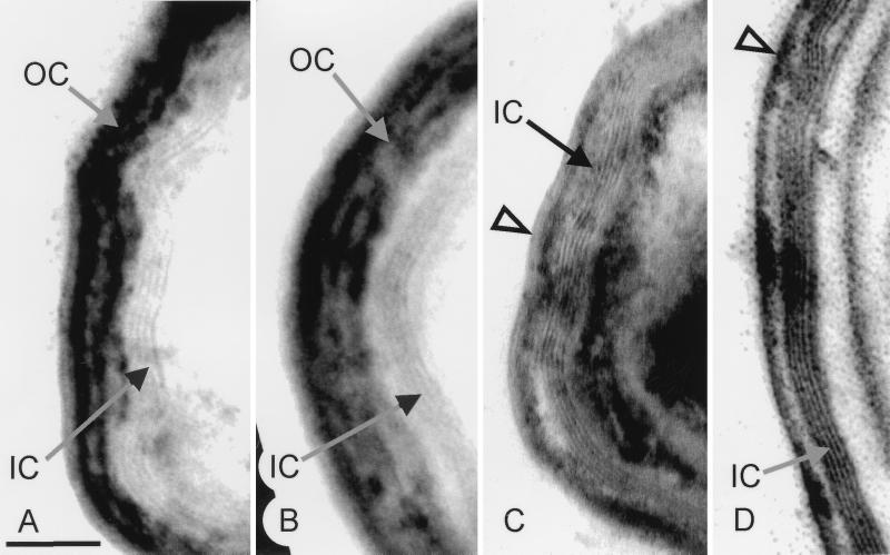
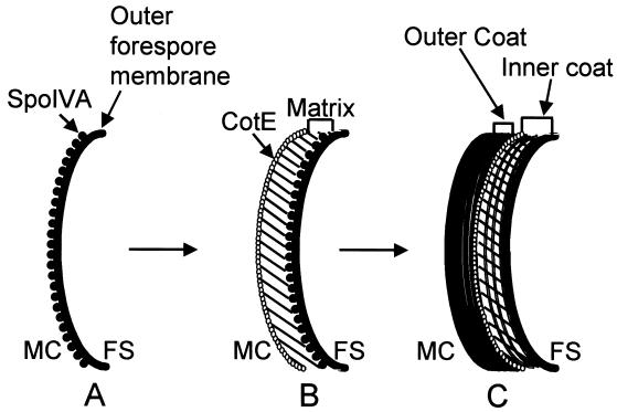
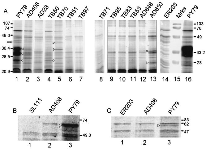
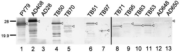
The Bacillus subtilis spore is encased in a resilient, multilayered proteinaceous shell, called the coat, that protects it from the environment. A 181-amino-acid coat protein called CotE assembles into the coat early in spore formation and plays a morphogenetic role in the assembly of the coat's outer layer. We have used a series of mutant alleles of cotE to identify regions involved in outer coat protein assembly. We found that the insertion of a 10-amino-acid epitope, between amino acids 178 and 179 of CotE, reduced or prevented the assembly of several spore coat proteins, including, most likely, CotG and CotB. The removal of 9 or 23 of the C-terminal-most amino acids resulted in an unusually thin outer coat from which a larger set of spore proteins was missing. In contrast, the removal of 37 amino acids from the C terminus, as well as other alterations between amino acids 4 and 160, resulted in the absence of a detectable outer coat but did not prevent localization of CotE to the forespore. These results indicate that changes in the C-terminal 23 amino acids of CotE and in the remainder of the protein have different consequences for outer coat protein assembly.


Figures

FIG. 1
FIG. 1
Electron microscopic analysis of wild-type and mutant spore coats. Arcs of wild-type (A), AD408 (B), TB50 (C), and TB70 (D) spore coats. The open arrowheads indicate thin remnants of the outer coat. Bar, 100 nm. IC, inner coat; OC, outer coat.
FIG. 2
FIG. 2
Model of the stages of coat formation. (A) SpoIVA localizes to the mother cell side of the sporulation septum, of which only an arc is shown. (B) The precoat, consisting of the matrix and the layer of CotE, assembles at the forespore surface, under the direction of SpoIVA. (C) The inner coat proteins assemble into the matrix, and the outer coat proteins bind around the shell of CotE. FS, forespore; MC, mother cell.
FIG. 3
FIG. 3
Deletion constructs of CotE and resulting phenotypes. The diagram in the upper left of the figure indicates the cotE open reading frame (box) and its upstream sequences (dotted line). cotE mutant constructs used in this study are indicated below the diagram. The triangles indicate oligonucleotides used to generate the deletion mutant versions of cotE. Asterisks indicate the positions of point mutations in AD648 and AD650. To the right of each construct is the strain name. The third column indicates which amino acids have been deleted or altered in each construct. The fourth column indicates the coat layers detected by electron microscopy (EM). OC, outer coat; IC, inner coat. The rightmost column gives the degree of lysozyme resistance, relative to that of the wild type. cotE null mutant spores have 6% resistance compared to the wild type. The numbers in parentheses are standard errors of the means.
FIG. 4
FIG. 4
SDS-PAGE analysis of spore coat extracts. Coat protein extracts were electrophoresed on 15% polyacrylamide gels. Extracts in each lane were prepared from spores of the indicated strain. (A) Coomassie blue-stained acrylamide gel. Lane 15 contains markers. The open arrow indicates CotS and the triangles between lanes 1 and 2 indicate the positions of bands of about 36, 30, and 26 kDa, discussed in the text. The triangles between lanes 4 and 5 indicate the positions of YvdO (upper) and YveN (lower) and the diamond indicates YrbB. The diamond between lanes 12 and 13 indicates the 31-kDa band, and the triangle indicates the 25-kDa band. Molecular masses indicated on the left are for lanes 1 to 13, and those on the right are for lanes 14 to 16. (B) Upper region of an SDS-polyacrylamide gel showing the consequence of deletion of cotA in AD408. (C) An experiment in which CotA and CotB were successfully resolved. In panels B and C, triangles indicate the positions of CotB. Molecular masses are indicated in kilodaltons.
FIG. 5
FIG. 5
Western blot analysis of spore coat extracts. Coat protein extracts were electrophoresed on 15% polyacrylamide gels and transferred to polyvinylidene difluoride membranes. CotE was detected with anti-CotE antibodies. Extracts in each lane were prepared from spores from the indicated strain. The positions of CotE are indicated by triangles. Bands below CotE are likely to be degradation products. The difference in migration between CotE in spores from PY79 and AD408 and the similarity in mobility of PY79 and TB50 are due to the additional mass of the HA1 epitope. Molecular masses are indicated in kilodaltons.
FIG. 6
FIG. 6
Immunofluorescence localization of CotE. At hour 4 of sporulation, cells were fixed for immunofluorescence microscopy, treated with either anti-CotE antibodies (panels A to J) or anti-SpoIVA antibodies (panel K), followed by a secondary Cy-5-conjugated antibody and then with the DNA stain DAPI (4′,6′-diamidino-2-phenylindole). Cells were examined for antibody staining (upper portion of each panel) or DNA staining (lower portion of each panel). Solid arrowheads indicate the more brightly fluorescing forespore chromosomes and the arrows indicate the diffuse mother cell chromosomes (17). Open arrowheads indicate positions of CotE decoration. (A) PY79; (B) AD28; (C) AD18; (D) AD408; (E) TB50; (F) TB51; (G) TB53; (H) TB83; (I) AD907; (J and K) AD648. Bar, 3 μm.

Similar articles

Cited by

References

    1. Abe A, Koide H, Kohno T, Watabe K. A Bacillus subtilis spore coat polypeptide gene, cotS. Microbiology. 1995;141:1433–1442. - PubMed
    1. Aronson A I, Fitz-James P. Structure and morphogenesis of the bacterial spore coat. Bacteriol Rev. 1976;40:360–402. - PMC - PubMed
    1. Cutting S M, Vander Horn P B. Molecular biological methods for Bacillus. Chichester, United Kingdom: John Wiley & Sons Ltd.; 1990.
    1. Donovan W, Zheng L, Sandman K, Losick R. Genes encoding spore coat polypeptides from Bacillus subtilis. J Mol Biol. 1987;196:1–10. - PubMed
    1. Driks, A. Unpublished observations.

Publication types

LinkOut - more resources