. 1999 Dec;73(12):9756-63.
doi: 10.1128/JVI.73.12.9756-9763.1999.

The activation domain of herpesvirus saimiri R protein interacts with the TATA-binding protein

Affiliations

The activation domain of herpesvirus saimiri R protein interacts with the TATA-binding protein

K T Hall et al. J Virol. 1999 Dec.

Abstract

The herpesvirus saimiri open reading frame (ORF) 50 produces two transcripts. The first is spliced, contains a single intron, and is detected at early times during the productive cycle, whereas the second is expressed later and is produced from a promoter within the second exon. Analysis of their gene products has shown that they function as sequence specific transactivators. In this report, we demonstrate that the carboxy terminus of ORF 50b contains an activation domain which is essential for transactivation. This domain contains positionally conserved hydrophobic residues found in a number of activation domains, including the herpes simplex virus VP16 and the Epstein-Barr virus R proteins. Mutational analysis of this domain demonstrates that these conserved hydrophobic residues are essential for ORF 50 transactivation capability. Furthermore, this domain is required for the interaction between the ORF 50 proteins and the basal transcription factor TATA-binding protein.


Figures

FIG. 1
FIG. 1
Schematic representation of the carboxy-terminus deletion series of the ORF 50b protein. A series of 3′ mutants were constructed by PCR amplification and ligated into the eukaryotic expression vector pBKCMV to derive pBK50Δ1-5.
FIG. 2
FIG. 2
In vitro transcription-translation analysis of ORF 50 deletion series. Protein expression from pBK50b (lane 1), pBK50Δ1 (lane 2), pBK50Δ2 (lane 3), pBK50Δ3 (lane 4), pBK50Δ4 (lane 5), and pBK50Δ5 (lane 6) was analyzed by in vitro transcription-translation. Transcription was initiated from the bacteriophage T3 promoter situated upstream of the cloned ORF50b fragments in pBKCMV. The synthesized products (indicated by arrows) were then separated on a 12% polyacrylamide gel and detected by autoradiography. Sizes are indicated in kilodaltons.
FIG. 3
FIG. 3
Expression levels and subcellular localization of ORF 50b and 50Δ1 proteins. A polyclonal antiserum was raised against a portion of recombinant ORF 50 protein and used in immunofluorescence analysis of cells mock transfected (i), HVS infected (MOI of 1) (ii), and transiently transfected with pBK50b (iii) and pBK50Δ1 (iv).
FIG. 4
FIG. 4
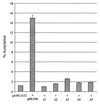
Analysis of the ORF 50b carboxy-terminus deletion series. OMK cell monolayers were transfected with 1 μg of pAWCAT2 or cotransfected 1 μg of each deletion plasmid, using DOTAP transfection reagent (Boehringer Mannheim) according to the manufacturer’s instructions. Cells were harvested at 48 h posttransfection, and cell extracts were assayed for CAT activity. Percentages of acetylation were calculated by the scintillation counting of the appropriate regions of the chromatography plate and are shown in graphical format; the variations between three replicated assays are indicated.
FIG. 5
FIG. 5
The carboxy-terminal ORF 50Δ1 construct produces a stable protein which binds to the ORF 50 response elements. (a) OMK cells were seeded at 106 cells per 35-mm-diameter petri dish and washed in labelling medium. Controls remained untransfected (lane 1) or were transfected with 2 μg of pBK50Δ1 (lane 2), pBK50 (lane 3) or infected with HVS (lane 4). The cells were incubated in labelling medium, harvested, and then lysed after 24 h. For each immunoprecipitation, 20 μl of the anti-ORF 50 polyclonal antibody was incubated with protein A-Sepharose beads for 16 h at 4°C. Immunoprecipitations were then performed with each cell lysate, using the anti-ORF 50 antibody. Beads were then pelleted, washed, and resuspended in Laemmli buffer; precipitated polypeptides were resolved on an SDS–12% polyacrylamide gel and analyzed by autoradiography. (b) Gel retardation assays were performed as previously described (53). Briefly, the ORF 50 response elements contained in a set of oligonucleotides were annealed and radiolabelled. These were incubated with nuclear extracts of untransfected OMK cells (lane 1) or cells transfected pBK50b (lane 2) and pBK50Δ1 (lane 3). The protein-nucleic acid complexes (indicated by arrow) were separated on a 5% polyacrylamide gel, run in 1% TBE buffer, and detected by autoradiography.
FIG. 6
FIG. 6
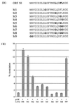
Mutational analysis of the ORF 50b transactivation domain. (a) The carboxy-terminal 14 amino acids contain a motif of positionally conserved hydrophobic amino acids homologous with the EBV R protein (boldface). A range of site directed mutations were constructed such that one or multiple conserved hydrophobic residues were replaced with a glycine residue (boldface). (b) OMK cell monolayers were transfected with 1 μg of pAWCAT2 or cotransfected with 1 μg of each 50b mutation plasmid, using DOTAP transfection reagent. Cells were harvested at 48 h posttransfection, and cell extracts were assayed for CAT activity. Percentages of acetylation were calculated by scintillation counting of the appropriate regions of the chromatography plate and are shown in graphical format; the variations between three replicated assays are indicated.
FIG. 7
FIG. 7
The ORF 50 carboxy-terminal mutation, 50M7, produces a stable protein which binds to the ORF 50 response elements. (a) Subcellular localization of ORF 50M7 protein, determined by immunofluorescence analysis of cells mock transfected (i) or transfected with pBK50M7 (ii). (b) OMK cells were seeded at 106 cells per 35-mm-diameter petri dish and washed in labelling medium. Controls remained untransfected (lane 1) or transfected with 2 μg of pBK50b (lane 2) or pBK50M7 (lane 3). The cells were incubated in labelling medium, harvested, and then lysed after 24 h. For each immunoprecipitation, 20 μl of the anti-ORF 50 polyclonal antibody was incubated with protein A-Sepharose beads for 16 h at 4°C. Immunoprecipitations were then performed with each cell lysate, using the anti-ORF 50 antibody. These samples were then pelleted, resuspended in Laemmli buffer, resolved on an SDS–12% polyacrylamide gel, and analyzed by autoradiography. (c) Gel retardation assays were performed as previously described (53). Briefly, the ORF 50 response elements contained in a set of oligonucleotides were annealed, radiolabelled and then incubated with nuclear extracts of untransfected OMK cells (lane 1) or cells transfected pBK50b (lane 2) and pBK50M7 (lane 3). The protein-nucleic acid complexes (indicated by the arrow) were separated on a 5% polyacrylamide gel, run in 1% TBE buffer, and detected by autoradiography.
FIG. 8
FIG. 8
The hydrophobic residues contained within the transactivation domain are required for the interaction with TBP. Immunoblot analysis was performed with the immunoprecipitation samples, untransfected control cells (lane 1) and cells transfected with 2 μg of pBK50b (lane 2) or pBK50M7 (lane 3). Polypeptides were resolved on an SDS–12% polyacrylamide gel and then transferred to nitrocellulose membranes. After transfer, the membranes were blocked, incubated with a 1/1,000 dilution of the anti-TFIID monoclonal antibody, washed, and incubated for 1 h at 37°C with a 1/1,000 dilution of anti-mouse immunoglobulin conjugated with horseradish peroxidase in blocking buffer. After five washes with PBS, the nitrocellulose membranes were developed, using enhanced chemiluminescence.
FIG. 9
FIG. 9
The ORF 50 transactivation domain is sufficient for the interaction with TBP. (a) The control GST alone (lane 1) and the GST-ORF 50 carboxy terminus fusion protein (lane 2) were expressed in E. coli DH5α and purified from crude lysates by incubation with glutathione-Sepharose 4B affinity beads. (b) The beads containing GST alone (lane 1) or the GST-ORF 50 carboxy terminus fusion protein (lane 2) were then incubated with OMK cell lysates. The beads were then pelleted, washed, resuspended in Laemmli buffer, and resolved on an SDS–12% polyacrylamide gel. The proteins were then transferred to nitrocellulose membranes by electroblotting and probed for TFIID.

Similar articles

Cited by

References

    1. Albrecht J C, Fleckenstein B. Structural organization of the conserved gene block of herpesvirus saimiri coding for DNA polymerase, glycoprotein B, and major DNA binding protein. Virology. 1990;174:533–542. - PubMed
    1. Albrecht J C, Nicholas J, Biller D, Cameron K R, Biesinger B, Newman C, Wittman S, Craxton M A, Coleman H, Fleckenstein B, Honess R W. Primary structure of the herpesvirus saimiri genome. J Virol. 1992;66:5047–5058. - PMC - PubMed
    1. Andrews N C, Faller D V. A rapid micropreparation technique for extraction of DNA-binding proteins from limiting numbers of mammalian cells. Nucleic Acids Res. 1991;19:2499. - PMC - PubMed
    1. Beisinger B, Muller-Fleckenstein I, Simmer B, Lang G, Wittmann S, Platzer E, Desrosiers R C, Fleckenstein B. Stable growth transformation of human T lymphocytes by herpesvirus saimiri. Proc Natl Acad Sci USA. 1992;89:3116–3119. - PMC - PubMed
    1. Bublot M, Manet E, Lequarre A S, Albrecht J C, Nicholas J, Fleckenstein B, Pastoret P P, Thiry E. Genetic relationships between bovine herpesvirus 4 and the gamma-herpesviruses Epstein-Barr and herpesvirus saimiri. Virology. 1992;190:654–665. - PubMed

Publication types

MeSH terms

LinkOut - more resources