. 1999 Dec;73(12):9764-72.
doi: 10.1128/JVI.73.12.9764-9772.1999.

Splicing regulatory elements within tat exon 2 of human immunodeficiency virus type 1 (HIV-1) are characteristic of group M but not group O HIV-1 strains

Affiliations

Splicing regulatory elements within tat exon 2 of human immunodeficiency virus type 1 (HIV-1) are characteristic of group M but not group O HIV-1 strains

P S Bilodeau et al. J Virol. 1999 Dec.

Abstract

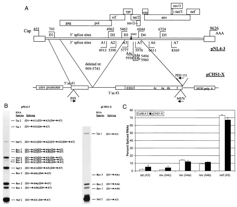
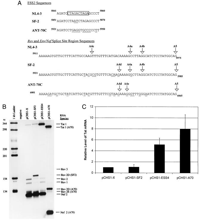
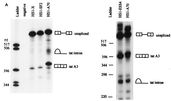
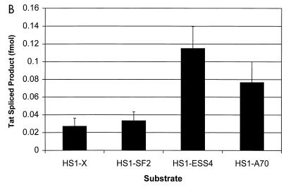
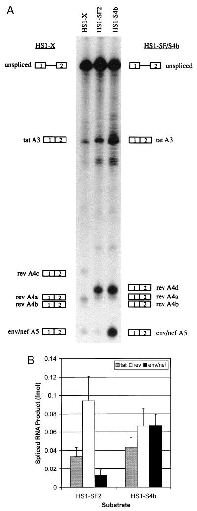
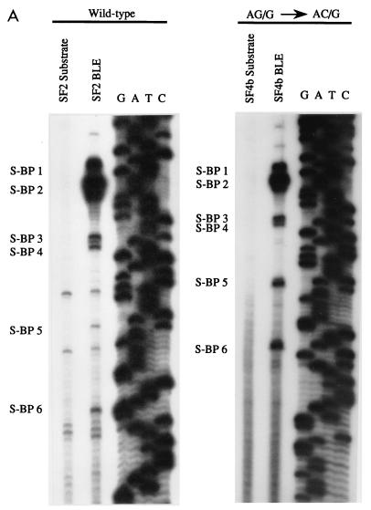
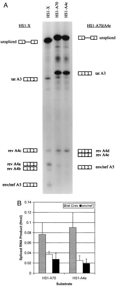
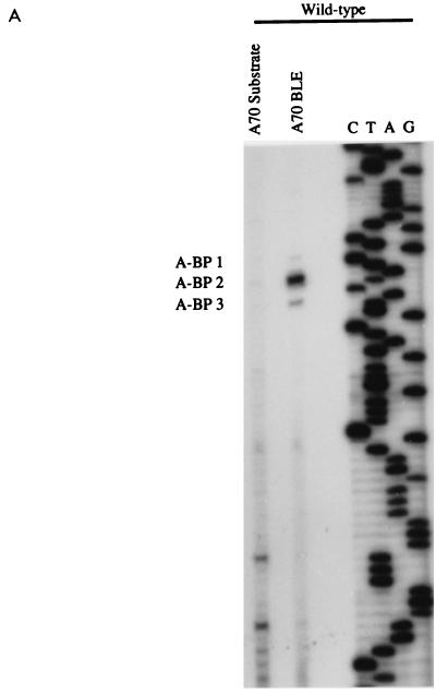
In the NL4-3 strain of human immunodeficiency virus type 1 (HIV-1), regulatory elements responsible for the relative efficiencies of alternative splicing at the tat, rev, and the env/nef 3' splice sites (A3 through A5) are contained within the region of tat exon 2 and its flanking sequences. Two elements affecting splicing of tat, rev, and env/nef mRNAs have been localized to this region. First, an exon splicing silencer (ESS2) in NL4-3, located approximately 70 nucleotides downstream from the 3' splice site used to generate tat mRNA, acts specifically to inhibit splicing at this splice site. Second, the A4b 3' splice site, which is the most downstream of the three rev 3' splice sites, also serves as an element inhibiting splicing at the env/nef 3' splice site A5. These elements are conserved in some but not all HIV-1 strains, and the effects of these sequence changes on splicing have been investigated in cell transfection and in vitro splicing assays. SF2, another clade B virus and member of the major (group M) viruses, has several sequence changes within ESS2 and uses a different rev 3' splice site. However, splicing is inhibited by the two elements similarly to NL4-3. As with the NL4-3 strain, the SF2 A4b AG dinucleotide overlaps an A5 branchpoint, and thus the inhibitory effect may result from competition of the same site for two different splicing factors. The sequence changes in ANT70C, a member of the highly divergent outlier (group O) viruses, are more extensive, and ESS2 activity in tat exon 2 is not present. Group O viruses also lack the rev 3' splice site A4b, which is conserved in all group M viruses. Mutagenesis of the most downstream rev 3' splice site of ANT70C does not increase splicing at A5, and all of the branchpoints are upstream of the two rev 3' splice sites. Thus, splicing regulatory elements in tat exon 2 which are characteristic of most group M HIV-1 strains are not present in group O HIV-1 strains.


Figures

FIG. 1
FIG. 1
RT-PCR analysis of multiply spliced HIV-mRNAs in cells transfected with wild-type and minigene NL4-3 constructs. (A) Structure of the NL4-3 HIV-1 genome. Locations of the known 5′ (D) and 3′ (A) splice sites (ss) and the ESS2 are shown. Boxes indicate open reading frames. Locations of the RNA initiation (Cap) and poly(A) site (AAA) are shown. Oligonucleotide primers used are indicated with arrows designating position and orientation. The construct pCHS1-X contains the indicated regions of pNL4-3. Location of the cytomegalovirus (cmv) promoter and human growth hormone poly(A) (HGH poly A) site are shown. (B) Representative polyacrylamide gel of products of RT-PCR using RNA from pNL4-3 and pCHS1-X. RT-PCR products of multiply spliced HIV-1 are designated according to the nomenclature of Purcell and Martin (15). “Splicing” indicates the 5′ and 3′ splice sites used to generate each RNA species. (C) Comparison of amounts of spliced products in genomic and minigene constructs based on multiple RT-PCR analyses. Shown are the percentages of products spliced at the following 3′ splice sites: tat (A3), rev (A4c), rev (A4a), rev (A4b), and env/nef (A5).
FIG. 2
FIG. 2
Comparison of tat, rev, and env/nef 3′ splice site usages in different HIV-1 strains by RT-PCR analysis. (A) Comparison of ESS2 sequences and the rev and env/nef splice site regions of HIV-1 strains NL4-3, SF2, and ANT70C. The core element within the ESS of pNL4-3 is boxed. The rev and env/nef splice sites are indicated by arrows, and the nucleotide numbers corresponding to GenBank sequences are shown. The sequence differences between SF-2 or ANT70C compared to the NL4-3 sequences are underlined. (B) RT-PCR analysis of spliced RNAs from mock-transfected HeLa cells or from HeLa cells transfected with pCHS1-X, pCHS1-SF2, pCHS1-ESS4, or pCHS1-A70. Rev 3 in the pCHS1-X lane is faint but is more detectable on longer exposures. The RT-PCR products from pCHS1-A70 migrate faster than those from NL4-3 and SF2 because the reverse primer (MS70) used for amplification of pCHS1-A70 products is located further upstream than the primer used for NL4-3 and SF2. (C) Relative amounts of spliced tat product. Multiple gels were quantitated, and the amounts of product are expressed relative to that of NL4-3 construct (pCHS1-X).
FIG. 3
FIG. 3
Comparison of tat 3′ splice site usage in different HIV-1 strains by in vitro splicing assays. (A) Denaturing PAGE of [32P]UTP-labeled HIV-1 substrates spliced in vitro. The positions of precursors, spliced products, and tat lariat products are indicated. On the left is a 6% gel comparing HS1-X, HS1-SF2, and HS1-A70; on the right is a 4% gel comparing HS1-ESS4 and HS1-A70. HS1-A70 spliced product migrates more slowly because the restriction site used to produce the linearized DNA template for transcription of RNA substrates is 7 nt further downstream than for HS1-X and HS1-SF2, resulting in longer RNA products. tat lariats migrate more slowly than the linear RNA species on 6% compared to 4% gels. (B) Quantitation of spliced tat RNA from the in vitro-spliced substrates shown in panel A. Multiple gels were quantitated, and the amounts of product spliced at the tat 3′ splice site (A3) were calculated based on uridine content of the RNA species.
FIG. 3
FIG. 3
Comparison of tat 3′ splice site usage in different HIV-1 strains by in vitro splicing assays. (A) Denaturing PAGE of [32P]UTP-labeled HIV-1 substrates spliced in vitro. The positions of precursors, spliced products, and tat lariat products are indicated. On the left is a 6% gel comparing HS1-X, HS1-SF2, and HS1-A70; on the right is a 4% gel comparing HS1-ESS4 and HS1-A70. HS1-A70 spliced product migrates more slowly because the restriction site used to produce the linearized DNA template for transcription of RNA substrates is 7 nt further downstream than for HS1-X and HS1-SF2, resulting in longer RNA products. tat lariats migrate more slowly than the linear RNA species on 6% compared to 4% gels. (B) Quantitation of spliced tat RNA from the in vitro-spliced substrates shown in panel A. Multiple gels were quantitated, and the amounts of product spliced at the tat 3′ splice site (A3) were calculated based on uridine content of the RNA species.
FIG. 4
FIG. 4
Mutagenesis of the rev A4b 3′ AG dinucleotide enhances splicing of the env/nef 3′ splice site in HS1-SF2. (A) Denaturing PAGE (6% gel) of [32P]UTP-labeled HS1-SF2 and rev A4b splice site mutant (HS1-SF4b) substrates spliced in vitro. Also shown is in vitro splicing of NL4-3 substrate HS1-X. HS1-SF4b is an AG-to-AC mutant at the A4b 3′ splice site. The positions of precursors and products spliced at the indicated splice sites are shown. (B) Quantitation of spliced tat, rev, and env/nef spliced products from the in vitro-spliced substrates shown in panel A. Multiple gels were quantitated, and the amounts of products spliced at the tat, rev, and env/nef 3′ splice sites were calculated based on of uridine content of the different RNA species.
FIG. 5
FIG. 5
Enhanced splicing at the env/nef A5 3′ splice site of HS1-SF2 correlates with increased use of downstream branchpoints. (A) Branchpoint location on BLEs of wild-type (HS1-SF2) and AG/G-to-AC/G mutant (HS1-SF4b) was carried out by primer extension analysis as described previously (22). Specific stops corresponding to branchpoints are shown. “Substrate” lanes indicate the same primer extension analysis carried out with unspliced precursor RNAs isolated from splicing gels. A dideoxy sequencing gel of the region was run simultaneously, and the results are shown. (B) Sequence alignment of NL4-3 and SF2 rev/env-nef splice site region. Locations of NL4-3 branchpoints sites have been described elsewhere (22). Branchpoints are indicated in bold underline type, and boxes represent BPSs. Splice sites are labeled by arrows.
FIG. 5
FIG. 5
Enhanced splicing at the env/nef A5 3′ splice site of HS1-SF2 correlates with increased use of downstream branchpoints. (A) Branchpoint location on BLEs of wild-type (HS1-SF2) and AG/G-to-AC/G mutant (HS1-SF4b) was carried out by primer extension analysis as described previously (22). Specific stops corresponding to branchpoints are shown. “Substrate” lanes indicate the same primer extension analysis carried out with unspliced precursor RNAs isolated from splicing gels. A dideoxy sequencing gel of the region was run simultaneously, and the results are shown. (B) Sequence alignment of NL4-3 and SF2 rev/env-nef splice site region. Locations of NL4-3 branchpoints sites have been described elsewhere (22). Branchpoints are indicated in bold underline type, and boxes represent BPSs. Splice sites are labeled by arrows.
FIG. 6
FIG. 6
Mutagenesis of rev 3′ splice site A4e of HS1-A70 does not increase splicing at the env/nef 3′ splice site A5. (A) Denaturing PAGE (6% gel) of [32P]UTP-labeled HS1-A70 and rev A4e splice site mutant (HS1-A4e) substrates spliced in vitro. Also shown is in vitro splicing of NL4-3 substrate HS1-X. HS1-A4e is an AG-to-AC mutant at the A4e 3′ splice site. The positions of precursors and products spliced at the indicated splice sites are shown. (B) Quantitation of spliced tat, rev, and env/nef spliced products from the in vitro-spliced substrates shown in panel A. Multiple gels were quantitated, and the amounts of products spliced at the tat, rev, and env/nef 3′ splice sites were calculated based on of uridine content of the different RNA species.
FIG. 7
FIG. 7
All detectable branchpoints in HS1-A70 are upstream of the rev 3′ splice sites. (A) Branchpoint analysis of BLEs from splicing reactions using the HS1-A70 substrate. (B) ANT70C sequence, location of the 3′ splice sites, and locations of the branchpoints (underlined).
FIG. 7
FIG. 7
All detectable branchpoints in HS1-A70 are upstream of the rev 3′ splice sites. (A) Branchpoint analysis of BLEs from splicing reactions using the HS1-A70 substrate. (B) ANT70C sequence, location of the 3′ splice sites, and locations of the branchpoints (underlined).

Similar articles

Cited by

References

    1. Adachi A, Gendelman H E, Koenig S, Folks T, Willey R, Rabson A, Martin M A. Production of acquired immunodeficiency syndrome-associated retrovirus in human and nonhuman cells transfected with an infectious molecular clone. J Virol. 1986;59:284–291. - PMC - PubMed
    1. Aiyar A, Leis J. Modification of the megaprimer method of PCR mutagenesis: improved amplification of the final product. BioTechniques. 1993;14:366–369. - PubMed
    1. Amendt B A, Hesslein D, Chang L-J, Stoltzfus C M. Presence of negative and positive cis-acting RNA splicing elements within and flanking the first tat coding exon of the human immunodeficiency virus type 1. Mol Cell Biol. 1994;14:3960–3970. - PMC - PubMed
    1. Amendt B A, Si Z-H, Stoltzfus C M. Presence of exon splicing silencers within human immunodeficiency virus type 1 tat exon 2 and tat/rev exon 3: evidence for inhibition mediated by cellular factors. Mol Cell Biol. 1995;15:4606–4615. - PMC - PubMed
    1. Bibollet-Ruche F, Loussert-Ajaka I, Simon F, Mboup S, Ngole E M, Saman E, Delaporte E, Peeters M. Genetic characterization of accessory genes from human immunodeficiency virus type 1 group O strains. AIDS Res Hum Retroviruses. 1998;14:951–961. - PubMed

Publication types

MeSH terms

Substances