. 1999 Dec;73(12):9816-26.
doi: 10.1128/JVI.73.12.9816-9826.1999.

The transcriptional switch of bacteriophage WPhi, a P2-related but heteroimmune coliphage

Affiliations

The transcriptional switch of bacteriophage WPhi, a P2-related but heteroimmune coliphage

T Liu et al. J Virol. 1999 Dec.

Abstract

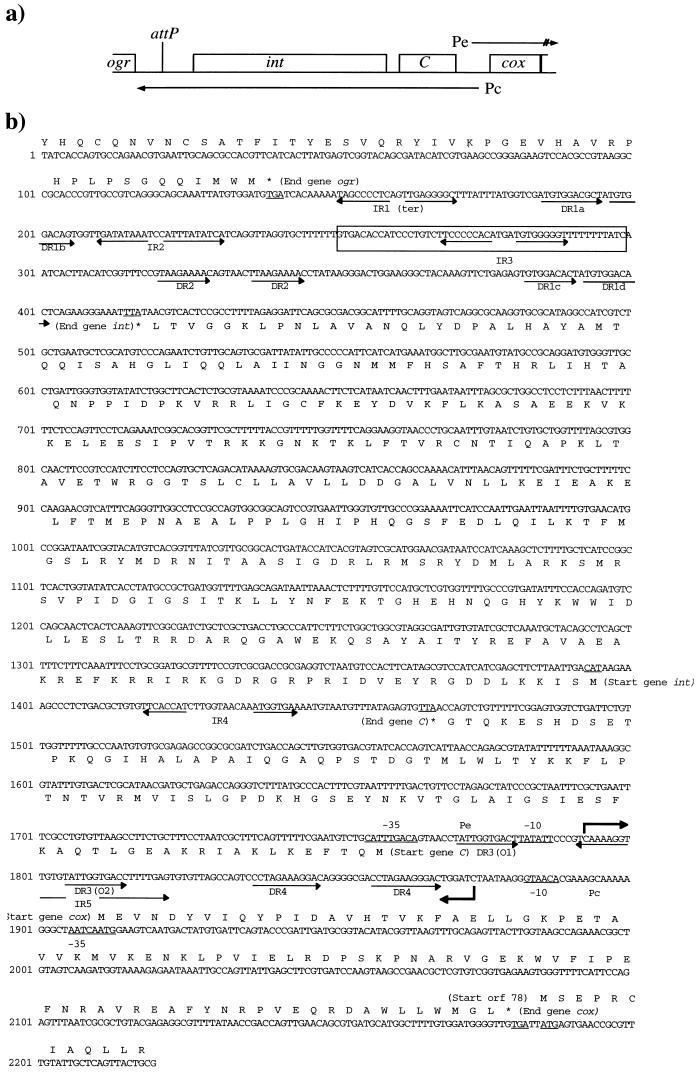
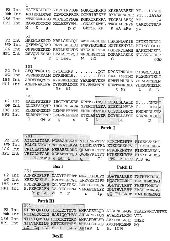
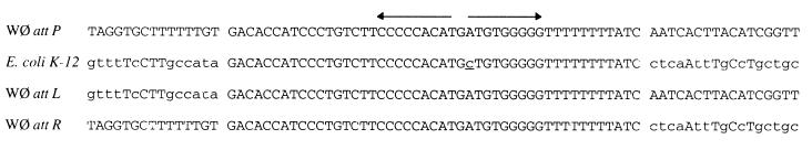
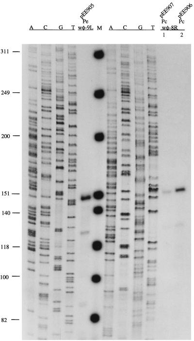
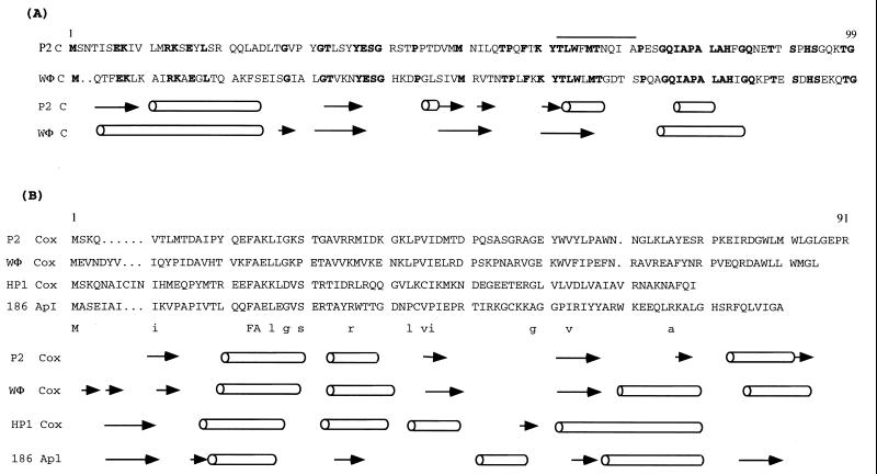
Phage WPhi is a member of the nonlambdoid P2 family of temperate phages. The DNA sequence of the whole early-control region and the int and attP region of phage WPhi has been determined. The phage integration site was located at 88.6 min of the Escherichia coli K-12 map, where a 47-nucleotide sequence was found to be identical in the host and phage genomes. The WPhi Int protein belongs to the Int family of site-specific recombinases, and it seems to have the same arm binding recognition sequence as P2 Int, but the core sequence differs. The transcriptional switch contains two face-to-face promoters, Pe and Pc, and two repressors, C and Cox, controlling Pe and Pc, respectively. The early Pe promoter was found to be much stronger than the Pc promoter. Furthermore, the Pe transcript was shown to interfere with Pc transcription. By site-directed mutagenesis, the binding site of the immunity repressor was located to two direct repeats spanning the Pe promoter. A point mutation in one or the other repeat does not affect repression by C, but when it is included in both, C has no effect on the Pe promoter. The Cox repressor efficiently blocks expression from the Pc promoter, but its DNA recognition sequence was not evident. Most members of the P2 family of phages are able to function as helpers for satellite phage P4, which lacks genes encoding structural proteins and packaging and lysis functions. In this work it is shown that P4 E, known to function as an antirepressor by binding to P2 C, also turns the transcriptional switch of WPhi from the lysogenic to the lytic mode. However, in contrast to P2 Cox, WPhi Cox is unable to activate the P4 Pll promoter.


Figures

FIG. 1
FIG. 1
(a) Schematic drawing of the transcriptional switch region of WΦ phage. The rightward transcript Pe is indicated on the top, and the leftward transcript Pc is shown on the bottom. attP is indicated, and the open reading frames named int, C, and cox are boxed. The coding region for ogr is represented by an open box. (b) Nucleotide sequence of the 2,220-bp region from phage WΦ DNA together with the deduced amino acid sequences of the open reading frames. ogr and orf78 are partly represented. Inverted repeats (IR) and direct repeats (DR) are indicated by back-to-back and rightward arrows, respectively. The core sequence of attP is indicated by a box. The predicated −10 and −35 sequences of the respective Pe and Pc promoter are underlined. The transcriptional start sites of Pe and Pc are indicated by bent arrows. The translational start and stop codens are also underlined.
FIG. 2
FIG. 2
Alignment of the Int amino acid sequences of four P2-related phages. Boxes I and II and patches I, II, and III are shaded. The identical residues are summarized in capital letters under the alignment, and those identical in three sequences are shown in lowercase letters.
FIG. 3
FIG. 3
Alignment of the core sequence and its flanking nucleotides of phage WΦ and its host E. coli K-12 together with the attL and attR sequence of WΦ prophage. The inverted repeat is indicated by back-to-back arrows. The mismatched nucleotide located in E. coli K-12 is underlined. Identical nucleotides are capitalized.
FIG. 4
FIG. 4
Autoradiograph of the primer extension products. Labelled primers wφ-9L and wφ-8R were annealed to RNA extracted from cells containing plasmid pEE905 (Pe) and pEE907 (Pc, lane 1) or pEE906 (Pc, lane 2). The corresponding sequence markers indicated as A, C, G, and T are positioned on the left side of the respective cDNA. Molecular markers (M) (in base pairs) are from end-labelled φX174 HinfI restriction fragments.
FIG. 5
FIG. 5
Amino acid sequence alignments and predicted secondary structures based on the conservation number from the Jpred program (14). Cylinders indicate helices, and arrows indicate β-sheets. (A) Repressor C of phage P2 and WΦ. The identical residues are shown in bold type. The proposed E binding site is indicated by a line above the P2 C sequence. (B) Cox/Apl proteins of phage P2, WΦ, HP1, and 186. The identical residues are summarized in capital letters under the alignment, and those identical in three sequences are shown in lowercase letters. The residue numbers above the sequence refer to P2 Cox.
FIG. 6
FIG. 6
Autoradiograph of CAT assay results from derepression of prophage WΦ by P4 E protein in the two-plasmid assay. The cells were prepared essentially as described previously (32). Samples were taken at different time points before and after isopropyl-β-d-thiogalactopyranoside (IPTG) induction of P4 E, as indicated at the bottom. Cell extracts contain 0.1 μg of protein in each assay mixture. −, control sample containing pKK232-8; +, control sample containing pSS32-1, which is the fully expressed P2 Pe promoter. CAT activity is normalized to that of P2 Pe promoter, which is set at 100.

Similar articles

Cited by

References

    1. Barreiro V, Haggård-Ljungquist E. Attachment sites for bacteriophage P2 on the Escherichia coli chromosome: DNA sequences, localization on the physical map, and detection of a P2-like remnant in E. coli K-12 derivatives. J Bacteriol. 1992;174:4086–4093. - PMC - PubMed
    1. Bertani G. Studies on lysogenesis. I. The mode of phage liberation by lysogenic Escherichia coli. J Bacteriol. 1951;62:293–300. - PMC - PubMed
    1. Bertani G, Chattoraj D K. Tandem pentuplication of a DNA segment in a derivative of bacteriophage P2: its use in the study of the mechanism of DNA annealing. Nucleic Acids Res. 1980;8:1339–1356. - PMC - PubMed
    1. Bertani G, Ljungquist E, Jagusztyn-Krynicka K, Jupp S. Defective particle assembly in wild type P2 bacteriophage and its correction by the lg mutation. J Gen Virol. 1978;38:251–261. - PubMed
    1. Bertani L E. Abortive induction of bacteriophage P2. Virology. 1968;36:87–103. - PubMed

Publication types

MeSH terms

Associated data

LinkOut - more resources