. 1999 Dec;73(12):9908-16.
doi: 10.1128/JVI.73.12.9908-9916.1999.

The residues between the two transformation effector sites of Epstein-Barr virus latent membrane protein 1 are not critical for B-lymphocyte growth transformation

Affiliations

The residues between the two transformation effector sites of Epstein-Barr virus latent membrane protein 1 are not critical for B-lymphocyte growth transformation

K M Izumi et al. J Virol. 1999 Dec.

Abstract

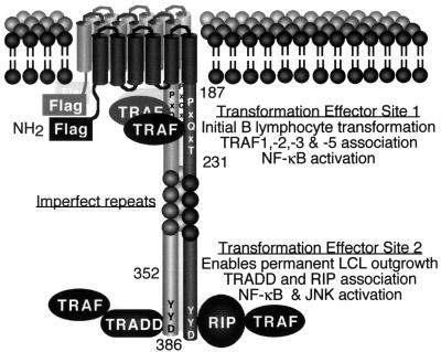
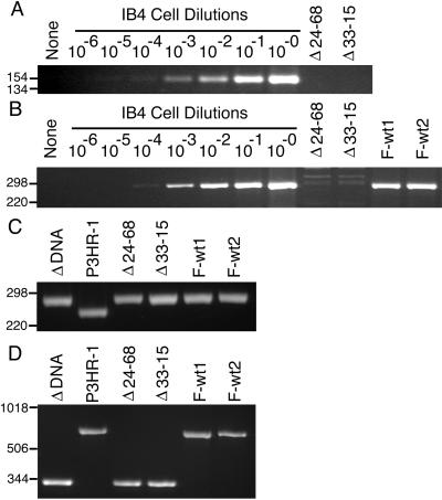
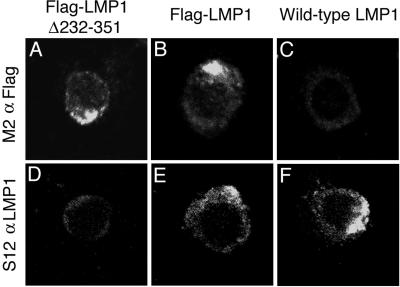
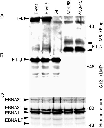
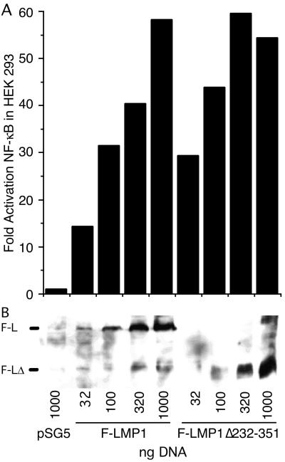
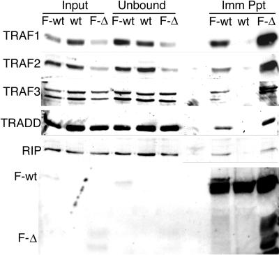
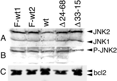
Epstein-Barr virus (EBV) latent membrane protein 1 (LMP1) is essential for EBV-mediated transformation of primary B lymphocytes. LMP1 spontaneously aggregates in the plasma membrane and enables two transformation effector sites (TES1 and TES2) within the 200-amino-acid cytoplasmic carboxyl terminus to constitutively engage the tumor necrosis factor receptor (TNFR)-associated factors TRAF1, TRAF2, TRAF3, and TRAF5 and the TNFR-associated death domain proteins TRADD and RIP, thereby activating NF-kappaB and c-Jun N-terminal kinase (JNK). To investigate the importance of the 60% of the LMP1 carboxyl terminus that lies between the TES1-TRAF and TES2-TRADD and -RIP binding sites, an EBV recombinant was made that contains a specific deletion of LMP1 codons 232 to 351. Surprisingly, the deletion mutant was similar to wild-type (wt) LMP1 EBV recombinants in its efficiency in transforming primary B lymphocytes into lymphoblastoid cell lines (LCLs). Mutant and wt EBV-transformed LCLs were similarly efficient in long-term outgrowth and in regrowth after endpoint dilution. Mutant and wt LMP1 proteins were also similar in their constitutive association with TRAF1, TRAF2, TRAF3, TRADD, and RIP. Mutant and wt EBV-transformed LCLs were similar in steady-state levels of Bcl2, JNK, and activated JNK proteins. The wt phenotype of recombinants with LMP1 codons 232 to 351 deleted further demarcates TES1 and TES2, underscores their central importance in B-lymphocyte growth transformation, and provides a new perspective on LMP1 sequence variation between TES1 and TES2.


Figures

FIG. 1
FIG. 1
Diagram of LMP1. The Flag epitope was introduced at the amino terminus (NH2). The positions of residues 187, 231, 352, and 386 are marked. LMP1 aggregates in the plasma membrane and constitutively associates with TRAFs, TRADD, and RIP. TES1 is located between residues 187 to 231 and aggregates TRAF1, -2, -3, and -5 to mediate NF-κB activation and initial B-lymphocyte growth transformation. TES2 is located between residues 352 and 386; aggregates RIP or TRADD, which associate with TRAFs to mediate high-level NF-κB and c-Jun N-terminal kinase (JNK) activation; and enables permanent LCL outgrowth.
FIG. 2
FIG. 2
PCR analyses of mutated LMP1 recombinant EBV-infected LCLs for LMP1 DNA. (A) PCR analysis for wt amino-terminal LMP1 DNA with the primers 5′-GAGGATGGAACACGACCTTGAGA-3′ and 5′-CTCCAGTCCAGTCACTCATAACG-3′. Lanes 8 to 2 are 104 IB4 cells (4 EBV DNA per cell) serially 10-fold diluted with 104 EBV-negative BJAB cells. After PCR amplification, DNAs were size separated in 3% agarose gels containing ethidium bromide. The endpoint dilution (lane 4) is the 10−4 dilution for a sensitivity of 4 copies of LMP1 DNA per 104 cells. No wt LMP1 DNA was detected in 104 Δ24–68 LCLs (lane 9) or Δ33–15 LCLs (lane 10) or in water (lane 1). Molecular markers (in base pairs) are indicated to the left of each panel. (B) PCR analysis for wild-type carboxyl-terminal LMP1 DNA as in panel A, except that the primers 5′-GACGGACCCCCACTCTGCTCTC-3′ and 5′-ATTGTGGAGGGCCTCCATCATTTC-3′ were used. (C) PCR analysis for Flag-LMP1 and wt LMP1 amino-terminal DNA as in panel A, except that the primers 5′-CACGCGTTACTCTGACGTAGCCG-3′ and 5′-CTCCAGTCCAGTCACTCATAACG-3′ were used. (D) PCR analysis for Flag-LMP1Δ232–351 deletion mutant and wt LMP1 carboxyl-terminal DNA as in panel A, except that the primers 5′-CTCTATTGGTTGATCTCCTTTGG-3′ and 5′-GCCTATGACATGGTAATGCCTAG-3′ were used.
FIG. 3
FIG. 3
Indirect immunofluorescent staining of methanol and acetone-fixed lymphoblastoid cell lines with M2 monoclonal antibody to Flag or S12 monoclonal antibody to the LMP1 carboxyl terminus. LMP1 spontaneously aggregates in the plasma membrane.
FIG. 4
FIG. 4
Protein expression in recombinant EBV-infected LCLs. (A) Western immunoblot analysis for Flag-LMP1. About 108 cells were Dounce homogenized in buffer containing 0.5% Brij 58, 100 mM NaCl, and 50 mM Tris (pH 7.2) and cleared by centrifugation. Flag-LMP1 was immunoprecipitated with M2 affinity gel (Sigma) for 6 h. Affinity gel was washed once, and precipitated proteins were detached with buffer containing SDS and 2-mercaptoethanol. About 10% of the immunoprecipitates were size separated in denaturing polyacrylamide gels, blotted to nitrocellulose filters, probed with M5 monoclonal antibody to Flag (Sigma) and peroxidase-conjugated secondary antibody to mouse immunoglobulin G (Amersham), and visualized by enhanced chemiluminescence (NEN Life Science). The position of Flag-LMP1 (F-L) is marked on the left, and the position of Flag-LMP1Δ232–351 (F-LΔ) is marked on the right. (B) Western immunoblot analysis for LMP1 carboxyl-terminal amino acids. About 5 × 10⁴ cells were lysed in buffer containing SDS and 2-mercaptoethanol and resolved in denaturing polyacrylamide gels. After Western transfer to nitrocellulose filters, LMP1 was detected as in panel A, except S12 monoclonal antibody was used. The position of Flag-LMP1 (F-L) or LMP1 (L) is marked on the left. (C) Western immunoblot analysis for EBV nuclear antigens (EBNA) leader protein (LP), EBNA1, EBNA2, and EBNA3C. The position of each protein is marked on the left. Analysis was done as in panel B, except that serum from a normal human donor and peroxidase-conjugated secondary antibody to human immunoglobulin G were used. In all panels, molecular mass markers (in kilodaltons) are marked on the right.
FIG. 5
FIG. 5
F-LMP1- and F-LMP1Δ232–351-expressing vectors activate NF-κB. (A) HEK293 cells (2.5 × 10⁵) were transfected (Qiagen Superfect) with the indicated amounts of F-LMP1- or F-LMP1Δ232–351-expressing vector or pSG5 vector control and with 350 ng of 3X-κB-L, a luciferase reporter with three NF-κB sites from human major histocompatibility class I and Fos minimal promoter and with 350 ng of pGK-β-gal DNA, a β-galactosidase-expressing vector used to monitor transfection efficiency. After 20 h at 37°C, cells were lysed in reference lysis buffer (Promega) and analyzed for luciferase activity (Promega) and β-galactosidase expression (Tropix) according to the manufacturer’s directions. (B) Western immunoblot analysis for F-LMP1 or F-LMP1Δ232–351. Equivalent amounts of protein from lysates prepared as described above were size separated and analyzed as described in the legend to Fig. 4A. The position of F-LMP1 or F-LMP1Δ232–351 is marked on the left.
FIG. 6
FIG. 6
Coimmunoprecipitation of TRAF1, TRAF2, TRAF3, TRADD, or RIP with F-LMP1 or F-LMP1Δ232–351. Proteins from LCLs (2.0 × 10⁸ cells) infected with F-LMP1, F-LMP1Δ232–351, or a wt LMP1 EBV recombinant were solubilized by Dounce disruption in 0.5% Brij 58, 100 mM NaCl, and 50 mM Tris (pH 7.2) and immunoprecipitated with M2 affinity gel (Sigma) to Flag. Precipitated proteins were Western immunoblot analyzed with antibodies to TRAF1, TRAF2, TRAF3, and TRADD from Santa Cruz Biotechnology, antibody to RIP from Pharmingen, or M5 antibody to Flag from Sigma. Input lanes represent unfractionated cell proteins, unbound lanes represent proteins not precipitated with M2 affinity gel, and Imm Ppt lanes represent immunoprecipitated proteins.
FIG. 7
FIG. 7
Western immunoblot analysis for c-Jun N-terminal kinase 1 and 2 (JNK1 and JNK2), phosphorylated JNK1 and JNK2 (P-JNK1 and P-JNK2), and Bcl2. LCLs (1.5 × 10⁶) transformed with F-LMP1, F-LMP1Δ232–351, or a wt LMP1 EBV recombinant were directly lysed in buffer containing SDS and 2-mercaptoethanol. Cell proteins were resolved in denaturing polyacrylamide gels, Western blotted to nitrocellulose filters, and probed with antibodies. (A) JNK1 and JNK2 are detected at similar levels in all LCLs with an antibody that recognizes JNK1 and JNK2 (New England Biolabs). (B) P-JNK2 but not P-JNK1 is detected at similar levels in all LCLs with an antibody that specifically recognizes P-JNK1 and P-JNK2 (New England Biolabs). (C) Bcl2 protein is detected at similar levels in all LCLs with a Bcl2-specific antibody (Santa Cruz Biotechnology).

Similar articles

Cited by

References

    1. Baichwal V R, Sugden B. Transformation of Balb 3T3 cells by the BNLF-1 gene of Epstein-Barr virus. Oncogene. 1988;2:461–467. - PubMed
    1. Brodeur S R, Cheng G, Baltimore D, Thorley L D. Localization of the major NF-κB-activating site and the sole TRAF3 binding site of LMP-1 defines two distinct signaling motifs. J Biol Chem. 1997;272:19777–19784. - PubMed
    1. Chen W G, Chen Y Y, Bacchi M M, Bacchi C E, Alvarenga M, Weiss L M. Genotyping of Epstein-Barr virus in Brazilian Burkitt’s lymphoma and reactive lymphoid tissue. Type A with a high prevalence of deletions within the latent membrane protein gene. Am J Pathol. 1996;148:17–23. - PMC - PubMed
    1. Cheng G, Baltimore D. TANK, a co-inducer with TRAF2 of TNF- and CD 40L-mediated NF-κB activation. Genes Dev. 1996;10:963–973. - PubMed
    1. Cohen J I, Wang F, Mannick J, Kieff E. Epstein-Barr virus nuclear protein 2 is a key determinant of lymphocyte transformation. Proc Natl Acad Sci USA. 1989;86:9558–9562. - PMC - PubMed

MeSH terms

Substances