. 1999 Dec;73(12):9917-27.
doi: 10.1128/JVI.73.12.9917-9927.1999.

Transcriptional up-regulation of the cyclin D2 gene and acquisition of new cyclin-dependent kinase partners in human T-cell leukemia virus type 1-infected cells

Affiliations

Transcriptional up-regulation of the cyclin D2 gene and acquisition of new cyclin-dependent kinase partners in human T-cell leukemia virus type 1-infected cells

F Santiago et al. J Virol. 1999 Dec.

Abstract

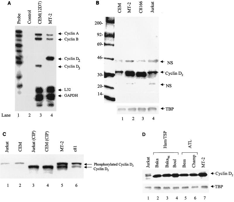
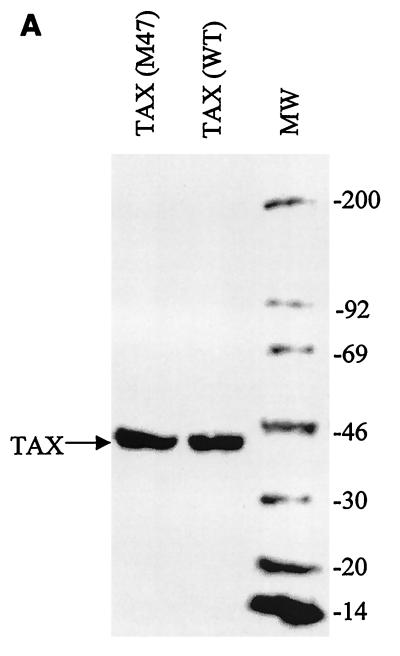
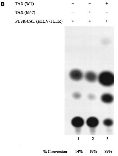
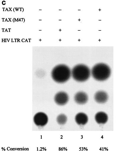
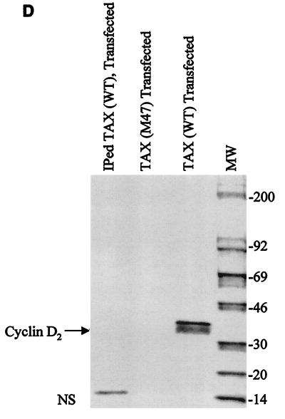
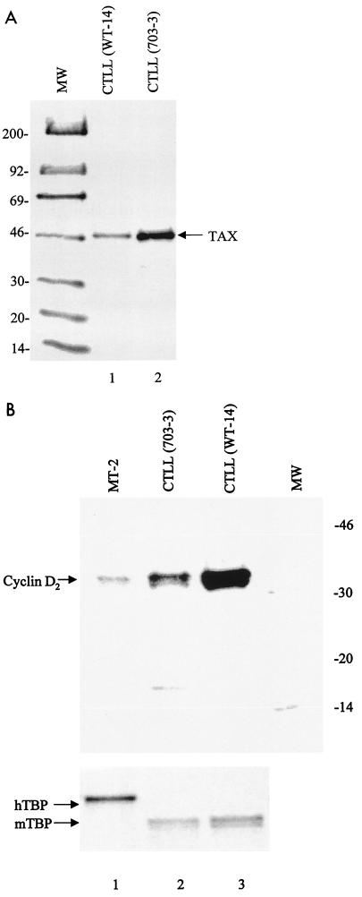
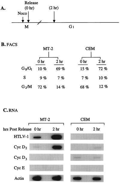
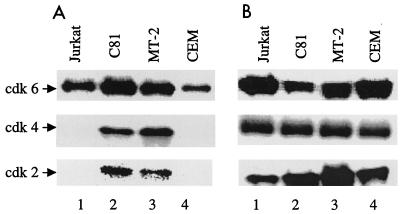
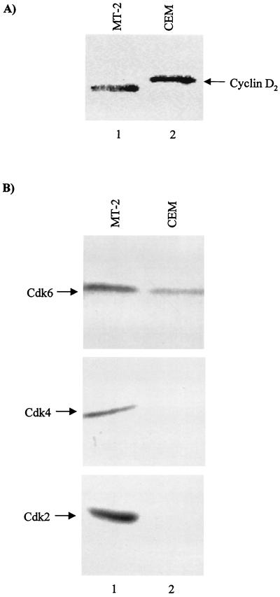
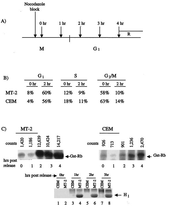
Human T-cell leukemia virus type 1 (HTLV-1) is the etiologic agent for adult T-cell leukemia/lymphoma (ATL) and HTLV-1-associated myelopathy/tropical spastic paraparesis. Tax(1) is a 40-kDa phosphoprotein, predominantly localized in the nucleus of the host cell, which functions to transactivate both viral and cellular promoters. It seems likely that HTLV-1, through expression of the viral regulatory protein Tax(1), provides some initial alteration in cell metabolism predisposing the development of ATL. Here, we demonstrate that HTLV-1 infection in T-cell lines and patient samples causes overexpression of an early G(1) cyclin, cyclin D2. The transcriptional up-regulation of the cyclin D2 gene is due to activation of Tax on the cyclin D2 gene. More important, we find that overexpression of cyclin D2 is accompanied by acquisition of new partners such as cyclin-dependent kinase 2 (cdk2), cdk4, and cdk6 in infected cells. This is in contrast to uninfected T cells, where cyclin D2 associates only with cdk6. Functional effects of these cyclin-cdk complexes in infected cells are shown by hyperphosphorylation of Rb and histone H1, indicators of active progression into S phase as well as changes in cellular chromatin and transcription machinery. These studies link HTLV-1 infection with changes of cellular cyclin gene expression, hence providing clues to development of T-cell leukemia.


Figures

FIG. 1
FIG. 1
Transcriptional activation of cyclin D2 in HTLV-1-infected cells. (A) Ten micrograms of RNA was used for hybridization with probes specific for cyclins A, B, C, D1, D2, D3, and A1. The human probe set used was human cyclin 1 from PharMingen. Following RNA preparation, hybridization, and digestion with RNases A and T1 as recommended by the manufacturer, protected fragments were separated on a 6% urea-polyacrylamide gel (Novex), dried, and exposed to a PhosphorImager cassette. Lane 1, 1/10 of the probe used for protection; lane 2, negative control sample hybridized with yeast tRNA; lanes 3 and 4, hybridization of uninfected (CEM) and HTLV-1-infected (MT-2) cells with the cyclin probes. Both L32 (cytoplasmic) and GAPDH (nuclear) RNA protections serve as internal controls in each lane. (B) Twenty-five micrograms of total cellular protein from uninfected (CEM and Jurkat) and infected (MT-2 and C8166) cells was prepared, separated by SDS-PAGE on a 4 to 20% gel, and blotted with anti-cyclin D2 polyclonal antibody or anti-TBP monoclonal antibody (generous gift from Nancy Thompson) (bottom). The antigen-antibody complex was further detected with 125I-protein G. The marker is a 14C-labeled Rainbow (high-molecular-weight) marker from Amersham; positions are indicated in kilodaltons. Cyclin D2 protein was seen at higher levels in HTLV-1-infected cells, as evident in lanes 2 and 3. Similar results have been obtained with two other cyclin D2 monoclonal antibodies, DCS-3 and DCS-5 (Neomarkers, Union City, Calif.). NS, nonspecific cross-reaction with cellular proteins. (C) Two hundred microgram of nuclear Jurkat or CEM extracts was treated with 100 U of CIP (Gibco/BRL catalog no. 18009-019), TCA precipitated, and run on a 6% Tricine-polyacrylamide (Novex) (lanes 3 and 4). Lanes 1, 2, 5, and 6 serve as controls (10 μg in each lane) for both phosphorylated and unphosphorylated cyclin D2. (D) Cellular extracts from four HTLV-1-infected patients, two with HAM/TSP and two with ATL, were processed and Western blotted with rabbit polyclonal anti-cyclin D2 antibody. All four samples were kept in culture for 4 to 5 months in the presence of exogenously added IL-2 (recombinant human IL-2; 200 U/ml; Boehringer Mannheim). A control TBP Western blot of the samples is shown at the bottom. The cells from ATL and HAM/TSP patients were not able to grow in the absence of IL-2, indicating that they are not fully transformed.
FIG. 2
FIG. 2
Activity of wild-type and mutant Tax protein on the endogenous cyclin D2 promoter. (A) Four hundred micrograms of purified and dialyzed wild-type (WT) and M47 Tax were run on an SDS–4 to 20% polyacrylamide gel and stained with Coomassie blue. MW, molecular weight markers (positions are indicated in kilodaltons on the right). (B) Two micrograms of each Tax protein and 3 μg of HTLV-1 reporter plasmid were transfected into CEM cells, and the cells were processed for CAT assay the next day (26). (C) As for panel B except that the reporter was HIV LTR-CAT and 200 ng of purified E. coli Tat was used as a control activator for this construct (lane 2). (D) Two micrograms of each Tax protein was transfected into 20 million CEM cells and processed 24 h later for Western blotting. Samples were lysed, and nuclear extracts were made as described in Materials and Methods, TCA precipitated, run on an SDS–4 to 20% polyacrylamide gel, and Western blotted with cyclin D2 antibody. In the IPed Tax (WT) lane (control), the wild-type Tax protein was immunoprecipitated with a cocktail of Tax monoclonal antibodies (Tab169, Tab170, Tab171, and Tab172) and pelleted in the presence of protein A+G-agarose, and the supernatant was used for transfection of CEM cells. NS, nonspecific reaction. (E) Recovery of Tax protein from the transfected cells. Details are as for panel D except that Western blotting was done with a cocktail of four anti-Tax monoclonal antibodies (1:500) and the antigen-antibody complex was detected with 125I-protein G (1:100; Amersham). Lane 1 and 2, controls where Tax was immunodepleted prior to transfection; lanes 3 and 4, nuclear extracts from transfected cells; lanes 5 and 6, 1/20 of the initial material used for transfection.
FIG. 2
FIG. 2
Activity of wild-type and mutant Tax protein on the endogenous cyclin D2 promoter. (A) Four hundred micrograms of purified and dialyzed wild-type (WT) and M47 Tax were run on an SDS–4 to 20% polyacrylamide gel and stained with Coomassie blue. MW, molecular weight markers (positions are indicated in kilodaltons on the right). (B) Two micrograms of each Tax protein and 3 μg of HTLV-1 reporter plasmid were transfected into CEM cells, and the cells were processed for CAT assay the next day (26). (C) As for panel B except that the reporter was HIV LTR-CAT and 200 ng of purified E. coli Tat was used as a control activator for this construct (lane 2). (D) Two micrograms of each Tax protein was transfected into 20 million CEM cells and processed 24 h later for Western blotting. Samples were lysed, and nuclear extracts were made as described in Materials and Methods, TCA precipitated, run on an SDS–4 to 20% polyacrylamide gel, and Western blotted with cyclin D2 antibody. In the IPed Tax (WT) lane (control), the wild-type Tax protein was immunoprecipitated with a cocktail of Tax monoclonal antibodies (Tab169, Tab170, Tab171, and Tab172) and pelleted in the presence of protein A+G-agarose, and the supernatant was used for transfection of CEM cells. NS, nonspecific reaction. (E) Recovery of Tax protein from the transfected cells. Details are as for panel D except that Western blotting was done with a cocktail of four anti-Tax monoclonal antibodies (1:500) and the antigen-antibody complex was detected with 125I-protein G (1:100; Amersham). Lane 1 and 2, controls where Tax was immunodepleted prior to transfection; lanes 3 and 4, nuclear extracts from transfected cells; lanes 5 and 6, 1/20 of the initial material used for transfection.
FIG. 3
FIG. 3
Effect of Tax on cyclin D2 expression. Mouse CTTL-2 (IL-2 dependent) cells were transfected with either wild-type or M47 Tax and selected for the ability to become IL-2 independent. Both cell types (described elsewhere [20]) were grown to mid-log phase of growth, and nuclear extracts were processed, run on an SDS–4 to 20% polyacrylamide gel, and Western blotted for either Tax (Tab172) or cyclin D2. (A) Wild-type (WT-14) and mutant (703-3) Tax Western blot analysis using 50 μg of extract. (B) Western blot analyses for mouse cyclin D2, using human antibody (top) and for both mouse and human TBP (hTBP), using polyclonal antibody (Santa Cruz) (bottom). Human and mouse cyclin D2 are more than 90% identical in primary sequence, and the human antibody cross-reacts with the mouse protein. CTLL (703-3) cells, which contain mutations at amino acids 319 and 320, show more than 80% reduction (wild type, 522,789 counts; 703-3, 6,325 counts) when quantitated on a PhosphorImager (Molecular Dynamics). MW lanes are as in Fig. 2A.
FIG. 4
FIG. 4
Endogenous promoter activities of HTLV-1 and early cyclin genes. MT-2 and CEM cells were blocked in low serum and nocodazole (Noco), washed the next day, and released. Samples were collected at time zero or at 2 h postrelease for RNA analysis. (A) Diagram of the experiment. (B) FACS analysis of both cell types, using propidium iodide DNA staining (FAST systems; Gaithersburg, Md.); (C) hybridization of 10 μg of total RNA, using HTLV-1 (nick translated sequence of HTLV-1 LTR, R region, +1 to +260) and cyclin D2, cyclin D3, cyclin E, and actin probes (1).
FIG. 5
FIG. 5
Various cdk partners of cyclin D2 in HTLV-1 infected cells. (A) Extracts from uninfected (Jurkat and CEM) and HTLV-1-infected (MT-2 and C8166) cells were used for immunoprecipitation with anti-cyclin D2 antibody and subsequently Western blotted with anti-cdk2, -4, and -6. Only cyclin D2 from HTLV-1-infected cells showed the presence of all three cdks in the complex. A number of antibodies specific to other cdks (cdk5, cdk7, cdk9, and cdc2) were used in cyclin D2 immunoprecipitation-Western blot assays and were found to be negative in HTLV-1-infected cells (data not shown). (B) 1/10 of the input cellular lysates used in immunoprecipitations.
FIG. 6
FIG. 6
Normalized concentrations of cyclin D2-associated complexes from infected and uninfected cells. A total of 500 μg of cellular proteins (MT-2 and CEM) was mixed with 50 μl of rabbit anti-human cyclin D2 antibody C-17 (Santa Cruz Biotechnology catalog no. sc-181) for immunoprecipitation and mixed for 12 to 14 h at 4°C; the next day, 150 μl of 30% protein G+A-agarose beads was added for 2 h, and the samples were pelleted, washed, and processed as in experiments represented in Fig. 5. (A) Western blot with anti-cyclin D2 antibody; (B) immunoprecipitation with anti-cyclin D2 antibody followed by Western blotting with anti-cdk2, -4, and -6 antibodies. Similar results were observed at higher concentrations of input (up to 10 mg) of MT-2 or CEM extract (data not shown).
FIG. 7
FIG. 7
Functional effects of cyclin D2-cdk partners from HTLV-1-infected cells. (A) Diagram of immunoprecipitations using anti-cyclin D2 antibody from both infected and uninfected cells treated with an M-phase blocker (nocodazole) and released. Following release, samples at various time points were processed and used for immunoprecipitations with cyclin D2 antibody. (B) FACS analysis of cells depicted in panel A following block and release with nocodazole. (C) Cyclin D2-immunoprecipitated complexes from infected and uninfected cells were washed and used in kinase assays with histone H1 and recombinant Rb proteins. Both cells traversed into the G1 phase following release, with higher kinase activity present in HTLV-1-infected cells when using Rb as a substrate (compare 2 to 4 h postrelease in MT-2 and CEM cells). However, only histone H1 (H1) was phosphorylated from HTLV-1-infected immunoprecipitates, implying that cdk2, which preferentially phosphorylated H1, is active in these cells (compare lanes 4 to 8).

Similar articles

Cited by

References

    1. Akagi T, Ono H, Shimotohno K. Expression of cell-cycle regulatory genes in HTLV-I infected T-cell lines: possible involvement of Tax1 in the altered expression of cyclin D2, p18Ink4 and p21Waf1/Cip1/Sdi1. Oncogene. 1996;12:1645–1652. - PubMed
    1. Arvanitakis L, Yaseen N, Sharma S. Latent membrane protein-1 induces cyclin D2 expression, pRb hyperphosphorylation, and loss of TGF-beta 1-mediated growth inhibition in EBV-positive B cells. J Immunol. 1995;155:1047–1056. - PubMed
    1. Bex F, Gaynor R B. Regulation of gene expression by HTLV-I Tax protein. Methods. 1998;16:83–94. - PubMed
    1. Blain S W, Montalvo E, Massague J. Differential interaction of the cyclin-dependent kinase (Cdk) inhibitor p27kip1 with cyclin A-Cdk2 and cyclin D2-Cdk4. J Biol Chem. 1997;272:25863–25872. - PubMed
    1. Bohan C A, Kashanchi F, Ensoli B, Buonaguro L, Boris-Lawrie K A, Brady J N. Analysis of Tat transactivation of human immunodeficiency virus transcription in vitro. Gene Expr. 1992;2:391–407. - PMC - PubMed

Publication types

LinkOut - more resources