

Hyperphosphorylation of the hepatitis C virus NS5A protein requires an active NS3 protease, NS4A, NS4B, and NS5A encoded on the same polyprotein
- PMID: 10559312
- PMCID: PMC113049
- DOI: 10.1128/JVI.73.12.9984-9991.1999
Hyperphosphorylation of the hepatitis C virus NS5A protein requires an active NS3 protease, NS4A, NS4B, and NS5A encoded on the same polyprotein
Abstract
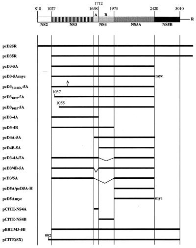
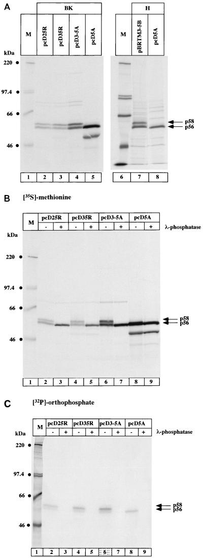
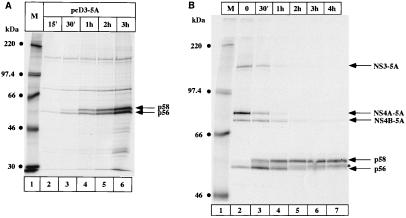
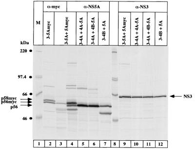
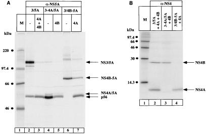
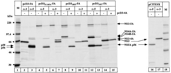
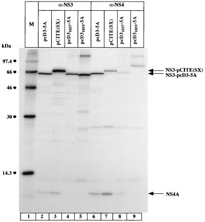
The nonstructural protein NS5A of hepatitis c virus (HCV) has been demonstrated to be a phosphoprotein with an apparent molecular mass of 56 kDa. In the presence of other viral proteins, p56 is converted into a slower-migrating form of NS5A (p58) by additional phosphorylation events. In this report, we show that the presence of NS3, NS4A, and NS4B together with NS5A is necessary and sufficient for the generation of the hyperphosphorylated form of NS5A (p58) and that all proteins must be encoded on the same polyprotein (in cis). Kinetic studies of NS5A synthesis and pulse-chase experiments demonstrate that fully processed NS5A is the substrate for the formation of p58 and that p56 is converted to p58. To investigate the role of NS3 in NS5A hyperphosphorylation, point and deletion mutations were introduced into NS3 in the context of a polyprotein containing the proteins from NS3 to NS5A. Mutation of the catalytic serine residue into alanine abolished protease activity of NS3 and resulted in total inhibition of NS5A hyperphosphorylation, even if polyprotein processing was allowed by addition of NS3 and NS4A in trans. The same result was obtained by deletion of the first 10 or 28 N-terminal amino acids of NS3, which are known to be important for the formation of a stable complex between NS3 and its cofactor NS4A. These data suggest that the formation of p58 is closely connected to HCV polyprotein processing events. Additional data obtained with NS3 containing the 34 C-terminal residues of NS2 provide evidence that in addition to NS3 protease activity the authentic N-terminal sequence is required for NS5A hyperphosphorylation.
Figures
Similar articles
-
Sequential Phosphorylation of the Hepatitis C Virus NS5A Protein Depends on NS3-Mediated Autocleavage between NS3 and NS4A.J Virol. 2020 Sep 15;94(19):e00420-20. doi: 10.1128/JVI.00420-20. Print 2020 Sep 15. J Virol. 2020. PMID: 32699091 Free PMC article.
-
Kinetic and structural analyses of hepatitis C virus polyprotein processing.J Virol. 1994 Aug;68(8):5045-55. doi: 10.1128/JVI.68.8.5045-5055.1994. J Virol. 1994. PMID: 8035505 Free PMC article.
-
Virus-specific cofactor requirement and chimeric hepatitis C virus/GB virus B nonstructural protein 3.J Virol. 2000 May;74(9):4291-301. doi: 10.1128/jvi.74.9.4291-4301.2000. J Virol. 2000. PMID: 10756044 Free PMC article.
-
Molecular virology of the hepatitis C virus.J Hepatol. 1999;31 Suppl 1:47-53. doi: 10.1016/s0168-8278(99)80374-2. J Hepatol. 1999. PMID: 10622560 Review.
-
The hepatitis C virus NS3 proteinase: structure and function of a zinc-containing serine proteinase.Antivir Ther. 1998;3(Suppl 3):99-109. Antivir Ther. 1998. PMID: 10726060 Review.
Cited by
-
Intragenic complementation of hepatitis C virus NS5A RNA replication-defective alleles.J Virol. 2013 Feb;87(4):2320-9. doi: 10.1128/JVI.02861-12. Epub 2012 Dec 12. J Virol. 2013. PMID: 23236071 Free PMC article.
-
Hepatitis C virus RNA replication is regulated by FKBP8 and Hsp90.EMBO J. 2006 Oct 18;25(20):5015-25. doi: 10.1038/sj.emboj.7601367. Epub 2006 Oct 5. EMBO J. 2006. PMID: 17024179 Free PMC article.
-
Human VAP-B is involved in hepatitis C virus replication through interaction with NS5A and NS5B.J Virol. 2005 Nov;79(21):13473-82. doi: 10.1128/JVI.79.21.13473-13482.2005. J Virol. 2005. PMID: 16227268 Free PMC article.
-
Mutational analysis of hepatitis C virus nonstructural protein 5A: potential role of differential phosphorylation in RNA replication and identification of a genetically flexible domain.J Virol. 2005 Mar;79(5):3187-94. doi: 10.1128/JVI.79.5.3187-3194.2005. J Virol. 2005. PMID: 15709040 Free PMC article.
-
Protease Inhibitors Block Multiple Functions of the NS3/4A Protease-Helicase during the Hepatitis C Virus Life Cycle.J Virol. 2015 May;89(10):5362-70. doi: 10.1128/JVI.03188-14. Epub 2015 Mar 4. J Virol. 2015. PMID: 25740995 Free PMC article.
References
-
- Afrikanova I, Fabbretti E, Miozzo M C, Burrone O R. Rotavirus NSP5 phosphorylation is up-regulated by interaction with NSP2. J Gen Virol. 1998;79:2679–2686. - PubMed
MeSH terms
Substances
LinkOut - more resources
Full Text Sources
Other Literature Sources
Research Materials
