. 1999 Dec;73(12):10000-9.
doi: 10.1128/JVI.73.12.10000-10009.1999.

Multiple effects of an anti-human immunodeficiency virus nucleocapsid inhibitor on virus morphology and replication

Affiliations

Multiple effects of an anti-human immunodeficiency virus nucleocapsid inhibitor on virus morphology and replication

L Berthoux et al. J Virol. 1999 Dec.

Abstract

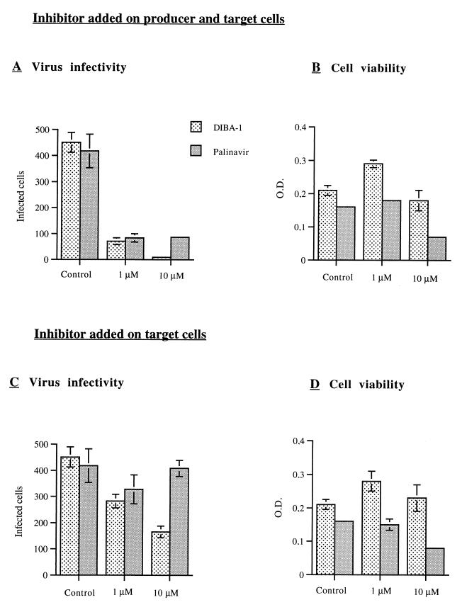
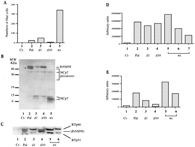
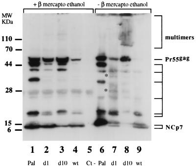
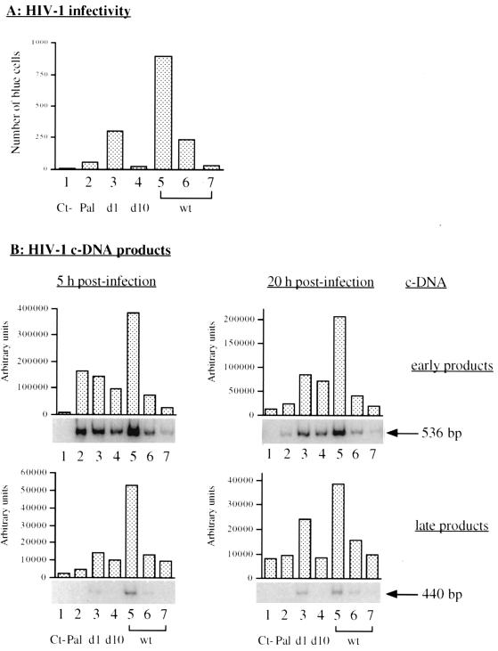
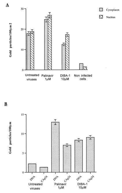
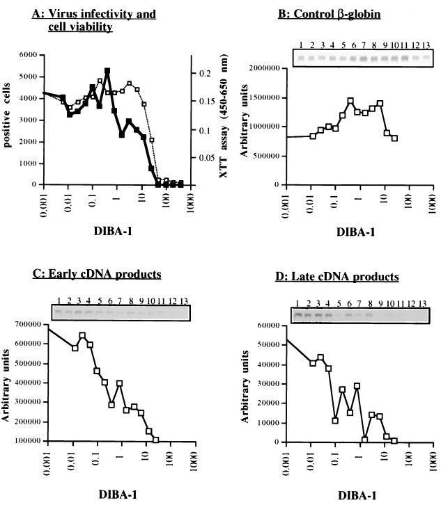
Human immunodeficiency virus type 1 nucleocapsid protein is a major structural component of the virion core and a key factor involved in proviral DNA synthesis and virus formation. 2,2'-Dithiobenzamides (DIBA-1) and related compounds that are inhibitors of NCp7 are thought to eject zinc ions from NCp7 zinc fingers, inhibiting the maturation of virion proteins. Here, we show that the presence of DIBA-1 at the time of virus formation causes morphological malformations of the virus and reduces proviral DNA synthesis. Thus, it seems that DIBA-1 is responsible for a "core-freezing effect," as shown by electron microscopy analyses. DIBA-1 can also directly interfere with the fate of the newly made proviral DNA in a manner independent of its effects on virion core formation. These data strongly suggest that nucleocapsid protein is a prime target for new compounds aimed at inhibiting human immunodeficiency virus and other retroviruses.


Figures

FIG. 1
FIG. 1
DIBA-1 and Palinavir act via distinct mechanisms. HeLa cells (3 × 10⁶) were transfected with pNL.EN and pCEL-E160 distributed into nine wells and processed as indicated in Materials and Methods. DIBA-1 or Palinavir was added at the indicated concentration 48 h prior to transfection or/and infection with HeLa-virus producer cells and CEMgfp target cells, or on CEMgfp target cells only, or with no inhibitor (control). (A and C) At 3 days after infection 100,000 CEMgfp cells were analyzed by FACS and GFP-positive cells were counted. (B and D) The viability of infected cells was determined prior to FACS analysis by XTT assay. Values correspond to an absorbance at 450 nm (after background substraction and with 650 nm as a reference) of 200 μl of cell supernatant, positively correlated to cell number and their metabolic activity. All results are given as the mean values of triplicates with the standard deviation.
FIG. 2
FIG. 2
Characterization of HIV-1 produced in the presence of DIBA-1 or Palinavir. HeLa cells were transfected with pNL4-3 and then distributed into 10-cm2 plates (see Materials and Methods). The next day, DIBA-1 or Palinavir was added, and 2 days later the supernatants were clarified. (A) Infectious titer. A 500-μl portion of supernatant was added to 105 HeLa and P4 cells plated the day before X-Gal staining was performed 24 h later. Values represent the total of blue cells per well. Columns: 1, negative control (supernatant from nontransfected cells); 2, 1 μM Palinavir; 3, 1 μM DIBA-1; 4, 10 μM DIBA-1; 5, untreated (positive control). (B) Maturation of virion proteins in the presence of DIBA-1 or Palinavir. Virions from 2 ml of supernatant were treated with SDS and 2-mercaptoethanol; viral proteins were then separated onto a 5 to 15% gradient acrylamide gel, blotted, and probed with an anti-NCp7 polyclonal antibody. Molecular weight markers are shown on the left, and viral proteins with the NC domain are indicated on the right. Lane 1, negative control Ct−; lane 2, 1 μM Palinavir; lane 3, 1 μM DIBA-1; lane 4, 10 μM DIBA-1; lane 5, untreated virus. (C) Presence of HIV-1 RT in DIBA-1- or Palinavir-treated virions. Samples were processed as in panel B, viral proteins were probed with polyclonal anti-NCp7 antibody, and membrane was stripped (but trace amounts of Pr55gag can still be seen) and reprobed with a polyclonal anti-RT antibody. Lane 1, negative control Ct−; lane 2, 1 μM Palinavir; lane 3, 1 μM DIBA-1; lane 4, 10 μM DIBA-1; lane 5, wild-type virus sample; lane 6, 25% of wild-type virus. (D) Virion genomic RNA content. Virion RNA corresponding to 5% of the supernatant was extracted, slot blotted, and probed with a DNA corresponding to a sequence in MA (see Materials and Methods). Slots were revealed by autoradiography, quantified by phosphorimaging, and expressed as arbitrary units (see Materials and Methods). Lane 1, negative control Ct−; lane 2, 1 μM Palinavir; lane 3, 1 μM DIBA-1; lane 4, 10 μM DIBA-1; lane 5, wild-type virus sample; lane 6, 25% of wild-type virus; lane 7, 15% of wild-type virus. (E) RT activity of DIBA-1- or Palinavir-treated virions. Portions (30 μl) of supernatant were subjected to exogenous RT assay as described before (4). Dots were revealed by autoradiography, quantified by phosphorimaging, and expressed as arbitrary units (see text). Lane 1, negative control Ct−; lane 2, 1 μM Palinavir; lane 3, 1 μM DIBA-1; lane 4, 10 μM DIBA-1; lane 5, wild-type virus sample; lane 6, 50% of wild-type virus.
FIG. 3
FIG. 3
Formation of disulfide bonds is induced by DIBA-1. Viruses were produced in the presence of inhibitor as described in Fig. 2. Proteins corresponding to 10 ml of supernatant were prepared for SDS-PAGE in reducing or nonreducing conditions and then resolved on a 5 to 15% polyacrylamide gel. Proteins were transferred to PVDF membranes and probed with a polyclonal anti-NCp7 antibody. Molecular weight markers are shown on the left. Lanes 1 and 6, 1 μM Palinavir; lanes 2 and 7, 1 μM DIBA-1; lanes 3 and 8, 10 μM DIBA-1; lanes 4 and 9, wild-type virus sample; lane 5, negative control. Note the formation of viral protein multimers containing NCp7 (the asterisks indicate discrete species).
FIG. 4
FIG. 4
Structure of HIV-1 virions produced in the presence of DIBA-1 or Palinavir. HeLa cells transfected with pNL4.3 were cultured in the presence of DIBA-1 or Palinavir. After 2 days the cells were prepared for electron microscopy. Representative viral particles, from left to right, included wild-type virus, Palinavir (1 μM), and DIBA-1 (10 μM). For DIBA-1-treated HIV-1, note the two virion structures corresponding to an immature morphology with frequent discontinuous peripheral rings (small arrows) or highly condensed and excentric cores (the freezing-core effect of DIBA-1; see large arrows). Also note that virions produced in the presence of DIBA-1 are much smaller (see text). The bar is 100 nm for all pictures.
FIG. 5
FIG. 5
Fate of HIV-1 cDNA synthesized in the presence of DIBA-1 or Palinavir. For pNL4.3 transfection and HeLa cell culture, see Materials and Methods. One day after transfection, inhibitor was added, and 2 days later supernatants were clarified and DNase treated. (A) Infectious titers. Portions (500 μl) of supernatant were added to 105 HeLa and P4 cells, and 24 h later X-Gal staining was carried out. Results are expressed as the number of blue cells per well. (B) PCR amplification of viral cDNA. Portions (500 μl) of supernatant were used to infect 2 × 10⁵ SupT1 cells for 5 h. Infections were also done with two dilutions (1/5 and 1/25) of untreated HIV-1. Cells were rinsed, and half were cultured for an additional 20 h. Nucleic acids were extracted, and [32P]dCTP-labelled PCR amplifications were carried out on 5% of each sample with primer pairs specific for early (corresponding to minus strand cDNA after the first transfer) and late (corresponding to double-stranded DNA after the second transfer) stages of the reverse transcription process (see panels). PCR DNA products were analyzed and quantified as indicated in Materials and Methods. Gels were autoradiographed (see results under the graphs) and submitted to phosphorimaging quantification (arbitrary units). Lane 1, negative control; lane 2, 1 μM Palinavir; lane 3, 1 μM DIBA-1; lane 4, 10 μM DIBA-1; lane 5, wild-type virus; lane 6, 20% of wild-type virus; lane 7, 4% of wild-type virus.
FIG. 6
FIG. 6
Effect of DIBA-1 or Palinavir on the localization of HIV-1 reverse transcription complexes. SupT1 cells were infected with HIV-1 produced in the presence of 1 μM palinavir or 10 μM DIBA-1. After 10 h cells were fixed and prepared for electron microscopy. The negative control is made up of noninfected cells. (A) Detection of early viral cDNA products. Sections were probed with an oligonucleotide complementary to the U5 sequence of genomic RNA. Gold particles were counted in the cytoplasm and nucleus areas of 30 randomly selected cells, and the densities are reported as numbers of gold particles per 100 μm2. (B) Nuclear membrane-associated viral cDNA and CAp24. Bars represent the density of gold particles corresponding to viral cDNA or CAp24 found at the nuclear envelope per 100 μm.
FIG. 7
FIG. 7
Inhibition of proviral DNA synthesis by DIBA-1. Single-cycle HIV-1 was produced by cotransfecting pNL4-3.EN− and pCEL/E160 in HeLa cells. The inhibitor-free virus preparation was used to infect CEMgfp cells cultured for 2 days in DIBA-1 (from 0 to 400 μM), and cells were cultured for 3 more days. (A) CEMgfp cell viability was monitored by use of the XTT assay (□), and the number of GFP-positive cells was determined (■; values represent the number of positive cells of 106 cells) for different concentrations of DIBA-1 (x axis). (B to D) Nucleic acids were extracted from one-third of the cells and subjected to radiolabelled PCR amplification in the presence of [32P]dCTP with primers specific for the β-globin gene (B), early (C), and late (D) cDNA products. Autoradiographs are shown on the top of each panel. Lane 1, control HIV-1; lanes 2 to 13, twofold increase from 0.01 to 20 μM DIBA-1. Quantifications by phosphorimaging are reported in arbitrary units.

References

    1. Adachi A, Gendelman H E, Koenig S, Folks T, Willey R, Rabson A, Martin M A. Production of acquired immunodeficiency syndrome-associated retrovirus in human and nonhuman cells transfected with an infectious molecular clone. J Virol. 1986;59:284–291. - PMC - PubMed
    1. Aldovini A, Young R A. Mutations of RNA and protein sequences involved in human immunodeficiency virus type 1 packaging result in production of noninfectious virus. J Virol. 1990;64:1920–1926. - PMC - PubMed
    1. Auxilien S, Keith G, Le Grice S F J, Darlix J-L. Role of post-transcriptional modifications of primer tRNALys.3 in the fidelity and efficacy of plus strand DNA transfer during HIV-1 reverse transcription. J Biol Chem. 1999;274:4412–4420. - PubMed
    1. Berthoux L, Péchoux C, Ottmann M, Morel G, Darlix J L. Mutations in the N-terminal domain of human immunodeficiency virus type 1 nucleocapsid protein affect virion core structure and proviral DNA synthesis. J Virol. 1997;71:6973–6981. - PMC - PubMed
    1. Chen C, Okayama H. High efficiency transformation of mammalian cells by plasmid DNA. Mol Cell Biol. 1987;7:2745–2752. - PMC - PubMed

Publication types

MeSH terms