. 1999 Dec;73(12):10029-39.
doi: 10.1128/JVI.73.12.10029-10039.1999.

An epitope of the Semliki Forest virus fusion protein exposed during virus-membrane fusion

Affiliations

An epitope of the Semliki Forest virus fusion protein exposed during virus-membrane fusion

A Ahn et al. J Virol. 1999 Dec.

Abstract

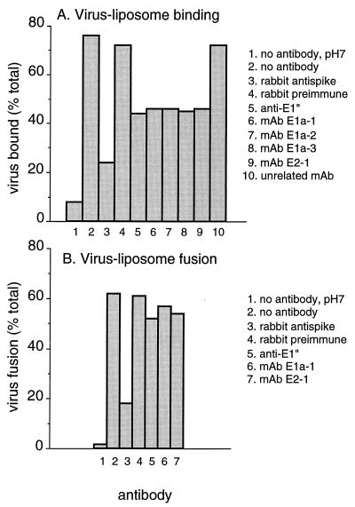
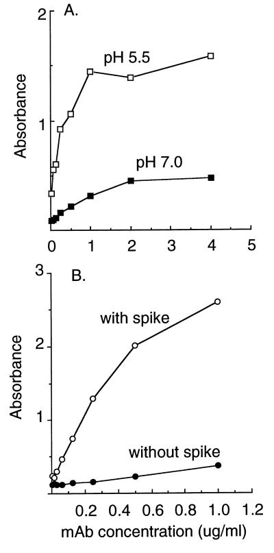
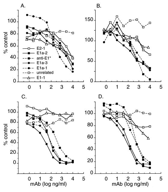
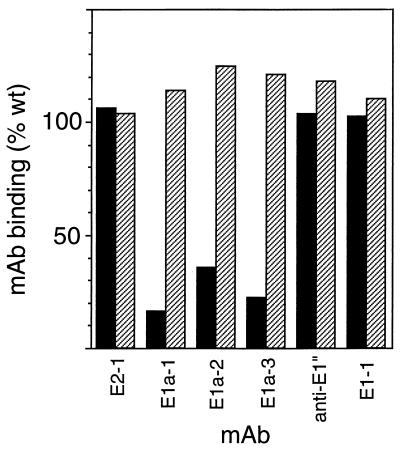
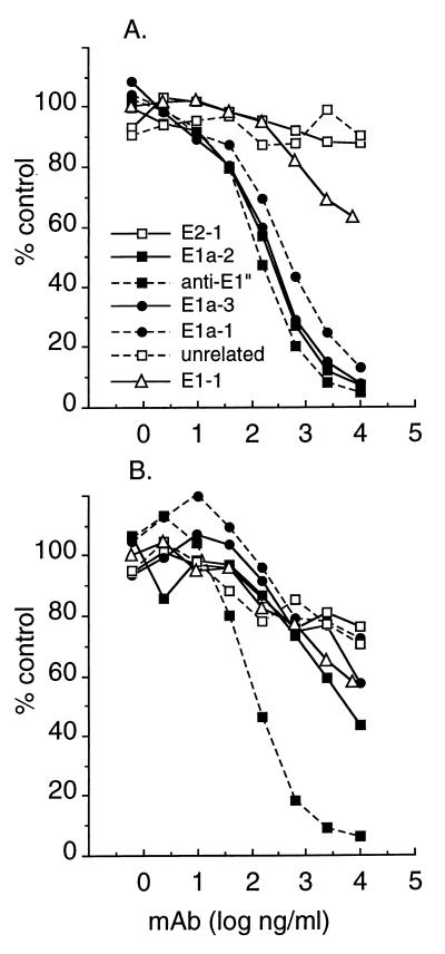
Semliki Forest virus (SFV) is an enveloped alphavirus that infects cells via a membrane fusion reaction triggered by acidic pH in the endocytic pathway. Fusion is mediated by the spike protein E1 subunit, an integral membrane protein that contains the viral fusion peptide and forms a stable homotrimer during fusion. We have characterized four monoclonal antibodies (MAbs) specific for the acid conformation of E1. These MAbs did not inhibit fusion, suggesting that they bind to an E1 region different from the fusion peptide. Competition analyses demonstrated that all four MAbs bound to spatially related sites on acid-treated virions or isolated spike proteins. To map the binding site, we selected for virus mutants resistant to one of the MAbs, E1a-1. One virus isolate, SFV 4-2, showed reduced binding of three acid-specific MAbs including E1a-1, while its binding of one acid-specific MAb as well as non-acid-specific MAbs to E1 and E2 was unchanged. The SFV 4-2 mutant was fully infectious, formed the E1 homotrimer, and had the wild-type pH dependence of infection. Sequence analysis demonstrated that the relevant mutation in SFV 4-2 was a change of E1 glycine 157 to arginine (G157R). Decreased binding of MAb E1a-1 was observed under a wide range of assay conditions, strongly suggesting that the E1 G157R mutation directly affects the MAb binding site. These data thus localize an E1 region that is normally hidden in the neutral pH structure and becomes exposed as part of the reorganization of the spike protein to its fusion-active conformation.


Figures

FIG. 1
FIG. 1
Functional effects of MAbs on SFV membrane attachment and fusion. (A) Virus-liposome attachment. [35S]methionine-[35S]cysteine-labeled wt SFV was mixed with liposomes and the indicated antibodies at final concentrations of 1.2 mM lipid and 50 μg of antibody per ml. The samples were preequilibrated for 3 to 5 min at 37°C, adjusted to pH 5.5 for 10 min at 37°C unless otherwise indicated, and then returned to neutral pH. Virus association with liposomes was quantitated by flotation on sucrose gradients (see Materials and Methods). Data shown are the average of two experiments. (B) Virus-membrane fusion. Pyrene-labeled wt SFV (0.6 μM) was mixed with unlabeled liposomes (200 μM) in the presence of 50 μg of the indicated antibodies per ml, preequilibrated for 3 to 5 min at 37°C, and adjusted to pH 5.5, and the final extent of fusion was measured after ∼30 s at 37°C by quantitating the decrease in the pyrene eximer peak by spectrofluorometry (see Materials and Methods). Treatment with the rabbit preimmune and anti-spike antibodies was performed by overnight incubation at 4°C followed by incubation as above. Data shown are the average of two or three experiments.
FIG. 2
FIG. 2
MAb E1a-1 binding to whole virus and isolated spike proteins. (A) Binding to whole virus. Gradient-purified wt SFV was pretreated at pH 5.5 or 7.0 for 5 min at 37°C as indicated, returned to neutral pH, and adjusted to 3 μg/ml. Virus was tethered to ELISA plates that were precoated with a MAb to the E2 spike subunit (E2-1). Biotin-conjugated MAb E1a-1 was added at the indicated concentrations and allowed to react with the virus for 1 h at 37°C. Biotinylated MAb binding was detected by incubation with streptavidin conjugated to alkaline phosphatase followed by incubation with substrate, each for 1 h at 37°C. Data shown are a representative example of four experiments. (B) Binding to adsorbed spike proteins. Using gradient-purified wt SFV, spike proteins were prepared by Triton X-114 phase separation and adsorbed directly to ELISA plates at 0.5 μg/ml. Biotin-conjugated MAb E1a-1 was added at the indicated concentrations and allowed to react with immobilized spike proteins for 1 h at 37°C. MAb binding was detected by incubation with streptavidan conjugated to alkaline phosphatase followed by incubation with substrate, each for 1 h at 37°C. The control consists of biotin-conjugated MAb E1a-1 binding in the absence of adsorbed spike proteins. Similar low absorbance was observed for binding of an unrelated MAb to wells containing spike protein. Data shown are a representative example of two experiments.
FIG. 3
FIG. 3
Antibody competition analysis. The ELISAs in Fig. 2 were used to analyze MAb binding to pH 5.5-treated intact wt virus (A and B) or purified wt spike proteins adsorbed to ELISA plates (C and D). The binding of biotin-conjugated MAb E1a-1 (A and C) or biotin-conjugated MAb anti-E1" (B and D) was quantitated at a constant biotin-MAb concentration of 100 ng/ml, in the presence of the indicated concentrations of competing, nonconjugated MAbs. Final binding results (in duplicate) were expressed as a percentage of the reactivity obtained with biotin-MAb in the absence of any competing MAb.
FIG. 4
FIG. 4
pH dependence of MAb E1a-1 binding to wt and mutant SFV. wt SFV and the 1-8 and 4-2 mutant virus isolates were diluted to a final titer of 108 PFU/ml in MES-saline-BSA buffer, and adjusted to the indicated pH for 5 min at 37°C. The samples were adjusted to neutral pH and 0.5% Triton X-100 and incubated in ELISA plates that were precoated with saturating concentrations of MAb E1a-1 (1 μg/ml). Bound spike protein was detected by the addition of a rabbit polyclonal antibody to the virus spike and an alkaline phosphatase-conjugated second antibody. Data shown are a representative example of four experiments.
FIG. 5
FIG. 5
MAb binding to isolated mutant spike proteins. wt virus, SFV 1-8, and SFV 4-2 were purified by gradient sedimentation, and spike proteins were isolated by Triton X-114 phase separation. Spike proteins were adsorbed directly to ELISA plates at 0.5 μg/ml and reacted with 100 ng of the indicated biotin-conjugated MAbs per ml. This concentration was within the linear range of MAb binding to wt spike proteins (Fig. 2B). MAb binding was detected by reacting with streptavidin conjugated to alkaline phosphatase, and mutant spike protein reactivity was expressed as percentage of wt binding. SFV 4-2 binding is shown as solid bars, and SFV 1-8 binding is shown as hatched bars. Data are a representative example of three experiments.
FIG. 6
FIG. 6
Antibody competition analysis of isolated mutant spike proteins. Purified spike proteins were prepared from SFV 1-8 (A) and SFV 4-2 (B) and adsorbed to ELISA plates, and the binding of biotin-conjugated MAb anti-E1" was quantitated in the presence of the indicated concentrations of competing, nonconjugated MAbs, all as in Fig. 3D. Final binding results (in duplicate) were expressed as a percentage of the reactivity obtained with biotin-MAb in the absence of any competing MAb.
FIG. 7
FIG. 7
Sensitivity of wt and mutant virus infection to inhibition by NH4Cl. BHK cells in 24-well trays were infected with 1 PFU of wt or mutant SFV per cell in the presence of the indicated concentration of NH4Cl for 90 min. Infection was then quantitated by determining the incorporation of [3H]uridine into viral RNA, in the presence of 20 mM NH4Cl to prevent secondary infection. Data shown are a representative example of two experiments.
FIG. 8
FIG. 8
MAb reactivity of isolated E1 homotrimers from wt and mutant SFV. [35S]methionine-[35S]cysteine-labeled wt SFV (A), SFV 1-8 (B), or SFV 4-2 (C) was mixed with liposomes (final concentration, 0.8 mM), adjusted to the indicated pH for 10 min at 37°C, and returned to neutral pH. One set of samples (pH 5 plus trypsin) was then treated with 200 μg of trypsin per ml in 1% Triton X-100 in PBS for 10 min at 37°C to digest E2, capsid, and any nontrimerized E1, and the digestion was terminated by the addition of trypsin inhibitor. All other samples were treated with premixed trypsin and trypsin inhibitor in 1% Triton X-100 in PBS. Antibody reactivity was analyzed by immunoprecipitation with the indicated antibodies followed by SDS-PAGE. rab indicates a rabbit polyclonal antibody against the SFV E1 and E2 subunits.

References

    1. Boere W A M, Harmsen T, Vinje J, Benaissa-Trouw B J, Kraaijeveld C A, Snippe H. Identification of distinct antigenic determinants on Semliki Forest virus by using monoclonal antibodies with different antiviral activities. J Virol. 1984;52:575–582. - PMC - PubMed
    1. Bordier C. Phase separation of integral membrane proteins in Triton X-114 solution. J Biol Chem. 1981;256:1604–1607. - PubMed
    1. Bron R, Wahlberg J M, Garoff H, Wilschut J. Membrane fusion of Semliki Forest virus in a model system: correlation between fusion kinetics and structural changes in the envelope glycoprotein. EMBO J. 1993;12:693–701. - PMC - PubMed
    1. Chatterjee, P. K., M. Vashishtha, and M. Kielian. Submitted for publication.
    1. Cheng R H, Kuhn R J, Olson N H, Rossman M G, Choi H-K, Smith T J, Baker T S. Nucleocapsid and glycoprotein organization in an enveloped virus. Cell. 1995;80:621–630. - PMC - PubMed

Publication types

LinkOut - more resources